Text-book of nervous diseases and psychiatry : for the use of students and practitioners of medicine / by Charles L. Dana.
- Charles Loomis Dana
- Date:
- 1904
Licence: Public Domain Mark
Credit: Text-book of nervous diseases and psychiatry : for the use of students and practitioners of medicine / by Charles L. Dana. Source: Wellcome Collection.
Provider: This material has been provided by the Augustus C. Long Health Sciences Library at Columbia University and Columbia University Libraries/Information Services, through the Medical Heritage Library. The original may be consulted at the the Augustus C. Long Health Sciences Library at Columbia University and Columbia University.
25/722 (page 3)
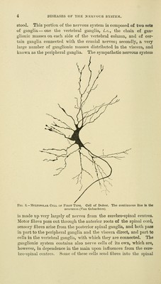
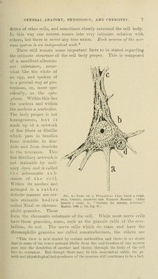
![are those adopted by the committee on anatomical nomenclature of the German Anatomical Society, and they have also been adoptea by a large number of writers ou neuro-anatora} . Beginning with the brain, we find that its particular subdivisions are based upon the embryological development of this organ. As will be shown in more detail later, the brain is developed out of three vesicles, known as the anterior, middle, and posterior vesicles (Fig. 1). The most anterior of these vesicles is the ^:>?'ost'«ce/^/taZo« or anterior brain ; the middle vesicle becomes the weseneephahni or inid-hrain, and the posterior vesicle develops into thQ rltombencejjha- Ion ov piosterior brain. Brain. fProsenceplialon ! (anterior brain). I' and I- Mesencephalon (middle brain).II Rhombencephalon (posterior brain). Ill and IV ^ 1. Telencephalon. ( 2. Diencepbalon. ■{ 3. Mesencephalon 4. Isthmus. 5. Metencephalon, 6. Myelencephalon. The anterior vesicle develops two secondary vesicles: the an terior portion of these, including the corpora striata, olfactory lobes, and the cerebral hemispheres, forms the telencep]ialon,V while the hinder portion of this vesicle, which includes the thalamus and mammary bodies, ioima the diencf^p/talon (1'). The middle vesicle i.>; the nie.sena-jjhulon, and it includes the corpora quadrigemiua and cerebral peduncles (II). The posterior vesicle is divided, from before backward, into three different parts: (1) the isthmus, which in- cludes the superior cerebellar peduncles and valve of Vieussens, and jjart of the cerebral peduncles; (2) the metenceplialon or /rind-brain. which includes the cerebrum and ]K)ns Varolii; and (o) the myelen. i-cphalon or after-brain, which includes the medulla oblongata. These different parts can be understood better by means of the .i.companying figure (Fig. 2), which represents in a schematic way the brain of a mammal (Edinger). They are intimately connected by strands of nerve fibres, and are connected closely also with the next portion of the nervous system, the spinal cord. The brain and spinal cord are spoki'u of as a cerebro-spinal axis, and this is in close relation with the p«n'ii)heral nervous system. This peripheral nervous system is composed of two portions— first, the cerebrij-spinal mixed nerves, whose origin, distribution, and relations are comitaratively easy to follow; and s('con<l, the gan- glionic or sympathetic nervous system. This has relations wliich are not so easily described, and wliich are as yet not wholly under-](https://iiif.wellcomecollection.org/image/b21224730_0025.jp2/full/800%2C/0/default.jpg)