Anatomy, descriptive and surgical / by Henry Gray ; the drawings by H.V. Carter ; with additional drawings in later editions.
- Henry Gray
- Date:
- 1897
Licence: Public Domain Mark
Credit: Anatomy, descriptive and surgical / by Henry Gray ; the drawings by H.V. Carter ; with additional drawings in later editions. Source: Wellcome Collection.
Provider: This material has been provided by the Francis A. Countway Library of Medicine, through the Medical Heritage Library. The original may be consulted at the Francis A. Countway Library of Medicine, Harvard Medical School.
69/1232 page 27
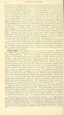
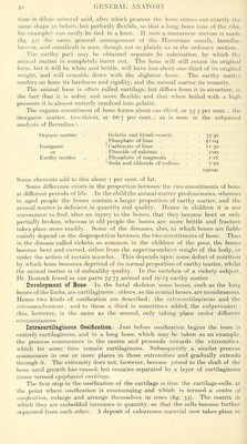
![the diploic tissue, the sicles of the canals being formed by a tlain lamella of bone, perforated here and there for the passage of branches from the adjacent cancelli. The same condition is also found in all cancellous tissue, the veins being enclosed and supported by osseous structure, and having exceedingly thin coats. When the bony structure is divided, the vessels remain patulous, and do not contract in the canals in which they are contained. Hence the occurrence of purulent absorption after amputation, in those cases where the stump becomes inflamed, and the cancellous tissue is infiltrated and bathed in pus. Lymphatic vessels, in addition to those found in the periosteum, have been traced, by Cruikshank, into the substance of bone, and Klein describes them as running in the Haversian canals. Nerves are distributed freely to the periosteum, and accompany the nutrient arteries into the interior of the bone. They are said by Kolliker to be most nun:ierous in the articular extremities of the long bones, in the vertebrae and the larger flat bones. Minute Anatomy.—The intimate structure of bone, which in all essential particulars is identical in the compact and cancellous tissue, is most easily studied in a transverse section from the compact wall of one of the long Fig. 28.—From a transverse section of the bones after maceration, such as is Jiaphysis of the humerus. Magnified 350 , . ^ o tmies. shown m ng. 28. If this is examined with a rather | ' ^ **•**'* low power the bone will be seen to 11^* ,-^, ? 'Aw* ^ i- * * be mapped out into a number of cir- ^/ * *.•' ^. ' .j^ * /^. cular districts : each one of which %^J^\ ]i- '^, «v A' consists of a central hole surrounded <.- f:- ^;ijx-v % :.'' _ I ' ' by a number of concentric rings. || <^,- . Jji'' 'Jt'S ^ *-** These districts are termed Haversian •*» ^ • --^ '^ w stjstems; the centralhole is a,n Haver- S W-W- ' J* ■ '«■« r sian canal, and the rings around are „ t^ ^Mi^' ** ^i \/ layers of bone-tissue arranged con- ^^' ; ' ^ ''fnm-' - *** » ' ^ ^^ centrically around the central canal, , ^j_^ ^ ' ' ' , \, -■«.'^^ H and termed lamella. Moreover, on ^^>^'.*'~*:^'''»/\ v^- \ ^ ^ closer examination, it will be found ' ' *^' '.'^ ' ,i^ thatbetween these lamellae, and there- ' 1*'' ' ', ■^ ' fore also arranged concentrically ^^ -^ifc' .!ggr . i^' ,/ around the central canal, are a num- t ^ .„ ,, . ,. ,.- a. Haversian canals, h. Lacunse, with their canahcnli la ber of little dark specks, the lacunce, the lamellte of these canals, c. Lacunre, of the intersti- T ,, , . 1 1 , T tial lamellfe. d. Others at the surface of the HaversiaM and that these lacunas are connected systems, with canaliculi given off from one side. with each other and with the central Haversian canal by a number of fine dark lines, which radiate like the spokes of a wheel and are called canalimli. All these structiu'es—the concentric lamellae, the lacvmae, and the canaliculi—may be seen in any single Haversian system, forming a circular district round a central, Haversian, canal. Between these circular sys- tems, filling in the irregular intervals which are left between them, are other lamellae, with their lacunae and canaliculi, running in various directions, but more or less curved (fig. 29). These are termed interstitial lamellge. Again, other lamellae, for the most part found on the surface of the bone, are arranged concentrically to the circumference of bone, constituting, as it were, a single Haversian system of the whole bone, of which the medullary cavity would represent the Haversian canal. These latter lamellae are termed circumferential, or by some authors primary ot fundamental lamellae, to distinguish them from those laid down around the axis of the Haversian canals, which are then termed secondary or sjiecial lamellas. The Haversian canals, seen as round holes in a transverse section of bone at or about the centre of each Haversian system, may be demonstrated to be true canals, if a longitudinal section is made, as in fig. 31. It will then be seen that](https://iiif.wellcomecollection.org/image/b21055129_0069.jp2/full/800%2C/0/default.jpg)

 No text description is available for this image
 No text description is available for this image
 No text description is available for this image