Anatomy, descriptive and surgical / by Henry Gray ; the drawings by H.V. Carter ; with additional drawings in later editions.
- Henry Gray
- Date:
- 1897
Licence: Public Domain Mark
Credit: Anatomy, descriptive and surgical / by Henry Gray ; the drawings by H.V. Carter ; with additional drawings in later editions. Source: Wellcome Collection.
Provider: This material has been provided by the Francis A. Countway Library of Medicine, through the Medical Heritage Library. The original may be consulted at the Francis A. Countway Library of Medicine, Harvard Medical School.
99/1232 page 57
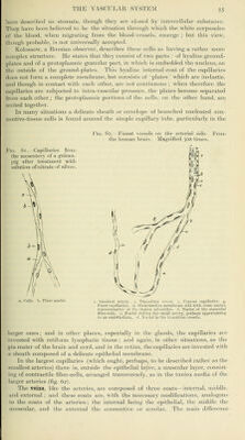
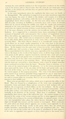
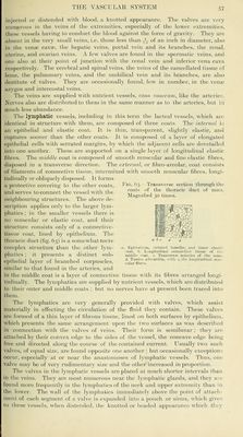
![injected or distended with blood, a knotted appearance. The valves are very numerous in the veins of the extremities, especially of the lower extremities, these vessels having to conduct the blood against the force of gravity. They are Absent in the very small veins, i.e. those less than J.,- of an inch in diameter, also in the venae cavae, the hepatic veins, portal vein and its branches, the renal, uterine, and ovarian veins. A few valves are found in the spermatic veins, and one also at their point of junction with the renal vein and inferior vena cava respectively. The cerebral and spinal veins, the veins of the cancellated tissue of bone, the pulmonary veins, and the umbilical vein and its branches, are also destitute of valves. They are occasionally found, few in number, in the venie iizygos and intercostal veins. The veins are supplied with nutrient vessels, vasa vasorum, like the arteries. Nerves also are distributed to them in the same manner as to the arteries, but in much less abundance. The lymphatic vessels, including in this term the lacteal vessels, which are identical in structure with them, are composed of three coats. The internal is an epithelial and elastic coat. It is thin, transparent, slightly elastic, and ruptures sooner than the other coats. It is composed of a layer of elongated epithelial cells wath serrated margins, by which the adjacent cells are dovetailed into one another. These are supported on a single layer of longitudinal elastic fibres. The middle coat is composed of smooth muscular and fine elastic fibres, disposed in a transverse direction. The external, or fibro-areolar, coat consists of filaments of connective tissue, intermixed with smooth muscular fibres, longi- tudinally or obliquely disposed. It forms a protective covering to the other coats, Fig. 63.-Transverse section through the and serves to connect the vessel with the coats of the thoracic duct of man. .... „, , T Magnined 30 times, neighbourmg structures. The above de- scription applies only to the larger lym- KrlTKlS^iSTj phatics; in the smaller vessels there is i'^ ^' '^^ no muscular or elastic coat, and their structure consists only of a connective- tissue coat, lined by epithelium. The thoracic duct (fig. 6^) is a somewhat more complex structure than the other lym- «. Epithelium, striated lamellas, a)id inner elastic ,,. .^ ^ Ti-ii uoat. I. Longitudinal connective tissue of the piiatlCS ; it presents a distinct sub- middle coat. «. Transverse muscles of the same. epthehal layer of branched corpuscles, ^^Tunk..^adventitia, with .^ similar to that found in the arteries, and in the middle coat is a layer of connective tissue with its fibres arranged longi- tudinally. The lymphatics are supplied by nutrient vessels, which are distributed to their outer and middle coats ; but no nerves have at present been traced into them. The lymphatics are very generally provided with valves, which assist materially in effecting the circulation of the fluid they contain. These valves are formed of a thin layer of fibrous tissue, lined on both surfaces by epithelium, which presents the same arrangement upon the two surfaces as was described in connection with the valves of veins. Their form is semilunar ; they are attached by their convex edge to the sides of the vessel, the concave edge being free and directed along the course of the contained current. Usually two such valves, of equal size, are found opposite one another; but occasionally exceptions occur, especially at or near the anastomoses of lymphatic vessels. Thus, one valve may be of very rudimentary size and the other increased in proportion. The valves in the lymphatic vessels are placed at much shorter intervals than in the veins. They are most numerous near the lymphatic glands, and. they are found more frequently in the lymphatics of the neck and upper extremity than in the lower. The wall of the lymphatics immediately above the point of attach- ment of each segment of a valve is expanded into a ]30uch or sinus, which give;^ to these vessels, when distended, the knotted or beaded appearance W'hich they](https://iiif.wellcomecollection.org/image/b21055129_0099.jp2/full/800%2C/0/default.jpg)
 No text description is available for this image
 No text description is available for this image
 No text description is available for this image