A report of microscopical & physiological researches into the nature of the agent or agents producing cholera / by T.R. Lewis and D.D. Cunningham.
- Timothy Richards Lewis
- Date:
- 1872
Licence: Public Domain Mark
Credit: A report of microscopical & physiological researches into the nature of the agent or agents producing cholera / by T.R. Lewis and D.D. Cunningham. Source: Wellcome Collection.
Provider: This material has been provided by The Royal College of Surgeons of England. The original may be consulted at The Royal College of Surgeons of England.
108/116 page 104
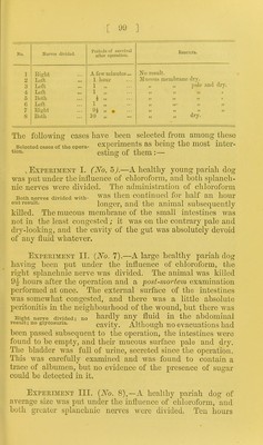
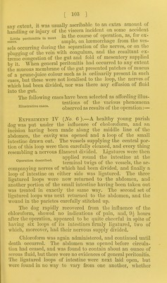
![[ 101. ] their nervous supply had been divided or not. They were all empty, and of a reddish brown ap- pearance internally, due to sanguine- This staining extended uninterruptedly from the highest ligatured loop to a little beyond the coecal extremity of the lowest one. Above the upper ligature, the mucous membrane of the gut was pale and somewhat more moist than in the ligatured loops. 1st case: no result. ous staining of the normal mucus Experiment Y {No. 8).—A strong healthy pariah dog was put under the influence of chloroform, and three loops of intestine were ligatured as in the preceding experiment, the nerves of the central loop being as before divided whilst those of the lateral loops were left intact. The intestine was then returned to the abdomen and the wound sewed up. After the lapse of an hour the abdomen was again opened, and the ligatured loops were examined. The vein of the central loop had become occluded with coagulum, which had caused extreme congestion veai^ extreme ccmgestion.tlie of the corresponding portion of the gut, but beyond this congestion no other difference existed between the central and the two lateral loops. Experiment VI {No. 9).—A large healthy pariah dog was put under the influence of chloroform, and the division of the nerves of a loop of intestine performed in the usual manner. Nine hours and a half subsequently the animal was kill- ed with chloroform, and the abdomen opened. The ligatured loop, the nerves of which had been divided, was distended like a sausage, and of an intense black colour. There was extreme extravasation along the lines of the vessels in the corresponding portion of mesentery, and the layers of the latter close to the intestine were widely separated by a wedge- . . nf shaped mass of tarry blood. The 3rd case: occlusion of tlie I i. l l *4-1 vein: sanguineous effusion. cavity of the gut WRS distended With black blood, and the tissues of its walls were infiltrated and thickened with similar fluid. This extreme congestion and extravasation had been caused by the complete occlusion^ of the vein by coagulum at the site of the section of the nerves. Experiment YII {No. 1G).—A small pariah dog nas put under the influence of chloroform, and the operation](https://iiif.wellcomecollection.org/image/b22355510_0110.jp2/full/800%2C/0/default.jpg)