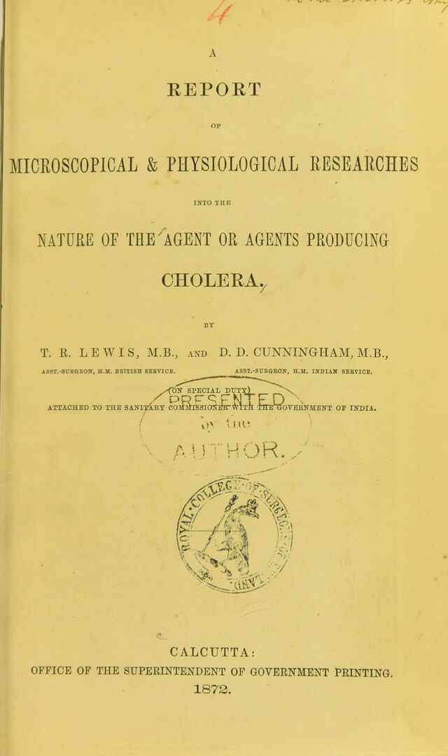
A report of microscopical & physiological researches into the nature of the agent or agents producing cholera / by T.R. Lewis and D.D. Cunningham.
- Timothy Richards Lewis
- Date:
- 1872
Licence: Public Domain Mark
Credit: A report of microscopical & physiological researches into the nature of the agent or agents producing cholera / by T.R. Lewis and D.D. Cunningham. Source: Wellcome Collection.
Provider: This material has been provided by The Royal College of Surgeons of England. The original may be consulted at The Royal College of Surgeons of England.
73/116 page 69
![[ 09 ] Experiment LXIV.—A large pariah dog was placed under chloroform, and an ounce of o/or^aaryUanrtn^discharge) the Same Solution as Used ill Expeii- 100 hours old: no effect. ment LVIII, &c., a hundred hours old, was injected into the right femoral vein. It continued drowsy for some time, vomited a large quantity of bilious matter, and by the next day was tolerably well, lho wound, however, had assumed an unhealthy, sloughy appearance, so the animal was killed forthwith. There was no peri- tonitis, the intestines were normal in every way, so were all the other viscera, thoracic and abdominal. The bladder was full. A wax-cell preparation of a drop of blood removed uo bacteria developed in f™ t'lc external iliac vein of the the blood. unwounded side, and a similar pre- paration of fluid pressed out of a mesenteric gland, were kept under observation for three days, during which period neither monads nor bacteria were seen in the former, but an abundance of white cells, whereas in the latter a few bacteria eventually appeared. Experiment LXV.—A large healthy pariah dog was placed under chloroform, and five ia^aelpcriment:Sd™aAafn 2i drachms of precisely the same fluid hours- as used in the last experiment were injected into its left femoral vein. After the operation it seemed to be much depressed, and vomited several times. The animal continued in this condition for two-and-a-half hours, when it died. A post-mortem examination was immediately made, and it was found that the small intestines, though very pale externally, were internally deeply congested, and the lumen congestion .of. villi and of the gut choked with a semi-fluid detachment of epithelium. slimy substance, consisting chiefly of detached epithelium, the individual cells being in a perfect state of preservation. Beneath this substance the villi were seen to be deeply congested, presenting a brush-like appear- ance. The stomach was healthy, and so was the large intes- tine. The mesenteric glands looked healthy, and so did the remainder of the abdominal viscera. There was no perito- nitis nor pleuritis, but there seemed to be some slight pericarditis. The lungs were collapsed and pale, and both sides of the heart contained fluid blood.](https://iiif.wellcomecollection.org/image/b22355510_0075.jp2/full/800%2C/0/default.jpg)