Symposium on Nuclear Sex / edited for the Organizing Committee by the secretary, D. Robertson Smith and [the chairman] William M. Davidson ; foreword by Robert Platt.
- Symposium on Nuclear Sex (1957 : King's College Hospital Medical School)
- Date:
- 1958
Licence: Attribution-NonCommercial 4.0 International (CC BY-NC 4.0)
Credit: Symposium on Nuclear Sex / edited for the Organizing Committee by the secretary, D. Robertson Smith and [the chairman] William M. Davidson ; foreword by Robert Platt. Source: Wellcome Collection.
43/216 page 21
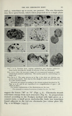
![THE SEX CHROMATIN BODY 21 and 4), sometimes up to seven, are present. The sex chromatin may show spiral forms, which when projected on to a single plane Figs, i to 6. Portions from amnion epithelium and chorion connective tissue nuclei showing the details of structure of the sex chromatin. Figs. 7a to с show the successive stages of a reconstructed amitosis or endo- mitosis from amniotic epithelium. The nuclear pairs in с each belong to a single cell. Figs. 8a to d. The same process as in Fig. 7 but these are chorion con¬ nective tissue nuclei. Note that in d the daughter nuclei are still connected by a fine nuclear strand. All nuclei are stained according to the thionin method described in (8). Figs. 3, 5, 7¿, and 80 are reproduced at a magnification of 3300 x, all others at 2600 X . For further explanations of the illustrations see the text. {These photographs are reproduced by permission of Academic Press Inc., New York.) suggest the letters M or S (Figs. 5, 6). Chromatin strands sometimes emerge from such structures (Fig. 6). In preparations stained according to the Feulgen method with light green as a counter-stain, a small green nucleolus-like body was sometimes found adjacent to the red sex chromatin [see colour plate III, Fig. 1С in Klinger (1957)].](https://iiif.wellcomecollection.org/image/b18020628_0044.JP2/full/800%2C/0/default.jpg)