Licence: Public Domain Mark
Credit: Auricular flutter / by William Thomas Ritchie. Source: Wellcome Collection.
Provider: This material has been provided by the Augustus C. Long Health Sciences Library at Columbia University and Columbia University Libraries/Information Services, through the Medical Heritage Library. The original may be consulted at the the Augustus C. Long Health Sciences Library at Columbia University and Columbia University.
165/206 page 113
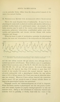
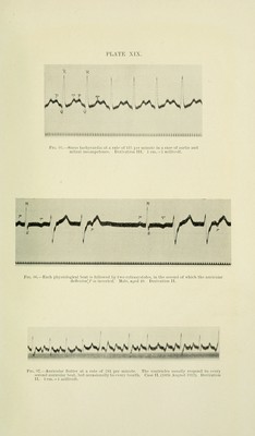
![c + a ill auiicular Hiitter dilTers from the sliai[»-iiointc(] sumniit of the wave f in a noiiiial ilivtlini. II. PllYSlOLOiilCAL lillVTHM WITH AUCELEIIATKJN (SiNUS TA<'IlVrAUDIA) This is the most frequent form of tachycardia. It may Ije due to })hysiological causes such as physical exertion and emotion influencing a perfectly healthy heart, or to pathological causes. As examples of the latter, mention may be made of the various acute exanthemata, lobar pneumonia, pulmonary tuberculosis, exophthalmic goitre, acute endo- carditis and myocarditis, and chronic valvular disease with cardiac dilatation and dropsy. When the heart, in spite of acceleration, maintains its physiological rhvthm, the onset and termination of the tachvcardia are both suradual, Fig. 03.—Sinu.s tachycardia, at the rate of 97 per minute, and puLsus alternans in a man, aged 57, with lieart failure. .Jugulo-carotid and brachial tracings. Tlie o-c interval i.s 024 second. and the rate seldom exceeds 140 per minute, even although there be marked myocardial enfeeblement. Thus in fifteen cases of exophthalmic goitre I found maximum rates of 83, 90, 97, 100. 107, 110,114,115, 117, 122, 128, 129, 131, 147 and 148 per mimite. In none of these cases was the As —Vs interval less than 0-13 second, or more than 0*17 second; it was usually 0'14 second—the normal interval. Again, in sub-acute endocarditis with a physiological rhythm the rate seldom rises to 150 or 160 per minute, but I have seen cases in which the latter rate was attained. Moreover, I have seldom observed the ventricular rate to exceed 135 per minute in cases of chronic valvular disease so long as the physiological rhythm was maintained. Polygraph records and electrocardiograms are of aid in diagnosis. When the heart's rhythm is physiological and the rate greatly acceler- ated, each carotid impulse in a jugular tracing is preceded by only one auricular wave. Owing to the curtailment of ventricular diastole each auricular wave is partially, or wholly, fused with the c wave of the 8](https://iiif.wellcomecollection.org/image/b21209388_0165.jp2/full/800%2C/0/default.jpg)
 No text description is available for this image
 No text description is available for this image
 No text description is available for this image