The fundus oculi, with an ophthalmoscopic atlas illustrating its physiological and pathological conditions.
- Frost, W. Adams.
- Date:
- 1896
Licence: Public Domain Mark
Credit: The fundus oculi, with an ophthalmoscopic atlas illustrating its physiological and pathological conditions. Source: Wellcome Collection.
Provider: This material has been provided by UCL Library Services. The original may be consulted at UCL (University College London)
283/446
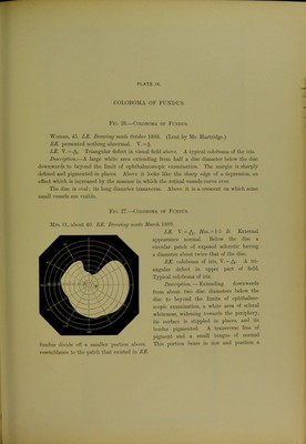
![COLOBOMA OF FUNDUS. Fig. 26.—Coloboma of Fundus. Woman, 45. LE. Dravring made October 1888. (Lent by Mr. Hartriclge.) RE. presented nothing abnormal. V. = f. LE. V. = Triangular defect in visual field above. A typical coloboma of the iris. DescriiJtion.—A large white area extending frona half a disc diameter below the disc downwards to beyond the limit of ophthalmoscopic examination. The margin is sharply defined and pigmented in places. Above it looks like the sharp edge of a depression, an effect which is increased by the manner in which the retinal vessels curve over. The disc is oval; its long diameter transverse. Above it is a crescent on which some small vesseLs are visible. Fig. 27.—Coloboma of Fundus. Mrs. 0., about 40. HE. Drcming made March 1889. LE. \. = ^, ffm. = l o D. External appearance normal. Below the disc a circular patch of exposed sclerotic having a diameter al)out twice that of the disc. BE. coloboma of iris, V. = A tri- angular defect in upper part of field. Typical coloboma of iris. Description. — Extending downwards from about two disc diameters below the disc to beyond the limits of ophthalmo- .scopic examination, a white area of scleral whiteness, widening towards the peripiiery, its surface is stippled in places, and its border pigmented. A transverse line of ])igment and a small tongue of normal fundus divide oil a smaller portion al)ove. This portion bears in size and position a resemVjlance to the patch that existed in HE.](https://iiif.wellcomecollection.org/image/b21271501_0283.jp2/full/800%2C/0/default.jpg)