The anatomy of the biting flies of the genus Stomoxys and Glossina / by G.M. Giles.
- Giles, George Michael, 1853-1916.
- Date:
- 1906
Licence: In copyright
Credit: The anatomy of the biting flies of the genus Stomoxys and Glossina / by G.M. Giles. Source: Wellcome Collection.
Provider: This material has been provided by The Royal College of Surgeons of England. The original may be consulted at The Royal College of Surgeons of England.
53/56
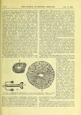
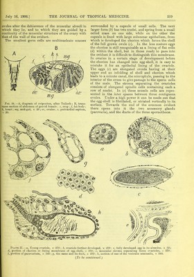
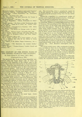
![Bassianum Laudum. “ Deoriginis et oausa pestis Patavinse.” Venetiis An., 1555, Id. “ Cura della Peste,” Yen., 1557. Th. Jordanus. “Pestis Phoenomena,” &c., Prancofurti, Wechelus, 1576. Massaria. “ De Peste,” Ven., 1597. Hier. Mercurialis. “ De peste preesertim de Veneya et Patavina,” Basel, 1577. Prosper Borgantius. “. De Peste,” Yen., 1565. Victor de Bongentibus. “Decern Problemata de Peste,” Ven., 1556. Georgius Agricola. “ De Peste in 1630,” Mediolanum, 1641. [24] Manget. “ Traite de la Peste, &c., Geneve, 1721,” pp. 214, 365, 551. [25] O’Meara. “ Oonquete de la Palestine,” 1799. Editee par Napoleon (without date). [26] Proust. “La defense de l’Europe contre la peste,” Paris, 1900. [27] Cabanes. Bull gin. de th(r.” November 30th, 1899. [28] J. Cantlie. Lancet, 1897, pp. 4-85 ; idem, 1897, p. 349, “Plague: How to Recognise, Prevent and Treat Plague,” London, 1900. [29] II Policlinico, 189S, p. 441. [30] G. Gaglio. Arcliivio per le scienze mediclie, vol. xxi., p. 341 ; A. Baldoni, Boll, della R. Acc. Medica di Roma, Ann. xxxi., Fasc. 1. [31] “Report of the Indian Plague Commission,” vol. v., p. 444. [32] Scheube. “ Die Krankeiten der warmen Lander,” Leipzig, 1900. [33] P. Manson. “ Tropical Diseases,” London : Cassell and Company, Ltd. THE ANATOMY OF THE BITING FLIES OF THE GENERA STOMOXYS AND GLOSSINA. By Lieut.-Colonel G. M. Giles, I.M.S. (Rtd.). (Continued from p. 219.) The parovaria (fig. 9) are two long, somewhat con- voluted tubes, the larger distal ends of which are attached to the branches of the oviduct near to the point, where they loose themselves in the stroma of the ovaries. They then pass backwards into the ovipositor, and then turn forwards again to their termination in the common oviduct. They have the same trabecular structure as the paragonia, but the trabeculae and the nuclei of their internodes are much larger, and a com- parison of fig. 9, Plate I., with fig. b, Plate II., shows that they also present resemblances in structure to that of the younger ovarioles. They appear to secrete a coagulable fluid similar to that of the paragonia. The remaining accessory structures are the spermothecae or receptaculae seminis, of which there are three, each of which (Plate II., fig. b.) consists of a dense chitinous sac supported in a cellular mass like an acorn in its cup. The chitinous membrane is fenestrated, as will be seen in fig. 2, and the ducts are supported by a spiral fibre somewhat like that of a trachea, but much coarser. Only one coitus takes place between the sexes, and in these sacs sufficient semen is stored on that occa- sion to last the life of the female. The ovipositor is quite of the usual type. Lieut. Tulloch describes it as follows :— “ The ovipositor consists of three cylindrical segments of thin chitin, which usually lie telescoped inside the abdomen. There is also a single external flap of dark chitin, which lies folded upon the ventral surface of the fly. When the ovipositor is extruded, by squeezing the abdomen, the recep- tacula and uterus are pulled down with it and can be seen through the transparent walls. The upper segment has three longitudinal rods of chitin, two dorsal and one ventral. The next is similar, but the last has the two dorsal plates only. The external flap, which is probably the ventral rib of the last segment, is roughly quadrilateral, and has no divergent prong-like processes arising from its free hinder border.” Within the ovipositor is a complicated system of circular and longitudinal striated muscles, which per- form the actions of protrusion and retraction. The male organs of Glossinct, as far as their histology is concerned, present no notable differences from those of Stomoxys, but according to Prof. Minchin, the para- gonia are more of the usual type, being distinct from each other throughout. Those of the female, on the other hand, depart entirely from the usual fly-type, being modified to meet the peculiar plan of reproduc- tion of these insects which give birth not to a multi- tude of eggs but to a very limited number of larvae. Owing to this, the common oviduct or uterus is of great size, and to expel the large full-grown larvae the ovipositor is provided with muscles, which, although on the same plan as those of Stomoxys, are so enormously developed that a section of this part of the body on a casual glance looks much like one of the thorax of an ordinary fly. Prof. Minchin’s description runs as follows :— “ The female genital organs differ considerably in appear- ance, according as they are in the gravid or non-gravid condition. In the course of my dissections I have only found one female in the latter state. In the later periods of gestation the condition of the female is obvious externally, but females which do not appear to be gravid are found on dissection to have a small larva in the uterus. “ The female organs (fig. 35) consist, like those of the male, of paired and unpaired portions. The former comprise the ovaries, the receptacula seminis and their ducts, and the Fig. 35.—The hinder segments of the abdomen with the female genital organs of Glossina in situ, dorsal view. rec. sem., recep- tacula seminis ; ov. r., ov. 1., right and left ovarioles ; d. rec., duct of the right receptaculum seminis; gl. ut., uterine glands (the greater number of these have been removed) ; c. d. gl., their common duct; retr. ut., retractor muscle of the uterus; Od., ovi- duct; TJt., uterus; L., hinder extremity of the larva, causing a bulge in the uterus ; pro. ut., protractor uteri, attached to the chitinous plate (Ch. 1); ni. vag., muscle (dilator vaginas?) passing from the vagina to the tergum of the seventh abdominal segment; m. v., muscle passing from the paired chitinous plate (Ch. 2) on each side of the vulva to the seventh tergum; Tag., vagina; V., vulva, the anterior margin of which is shown by a dotted line; An., anus; Ch. 1, Ch. 2, paired chitinous plates. (After Tulloch.)](https://iiif.wellcomecollection.org/image/b22416602_0055.jp2/full/800%2C/0/default.jpg)