Volume 1
Elements of anatomy / / edited by William Sharpey, Allen Thomson, and Edward Albert Schafer.
- Jones Quain
- Date:
- 1878
Licence: Public Domain Mark
Credit: Elements of anatomy / / edited by William Sharpey, Allen Thomson, and Edward Albert Schafer. Source: Wellcome Collection.
Provider: This material has been provided by Royal College of Physicians, London. The original may be consulted at Royal College of Physicians, London.
30/720 page 10
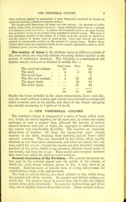
![are pierced by numerous small foramina for the passage of blood-ves- sels, one or two of whicb foramina situated near the middle of the pos- terior surface are much larger than the others. The arch consists of two symmetrical portions which spring, one on each side, fi'om the posterior surface of the body, and meet in the middle line behind. The anterior part of each half, rounded and nar- row, is called the pedicle; the posterior partis broad and flat, and is called the lamina (neurapophysis, Owen). The concavities on the upper and lower borders of the pedicles are named notches, and consti- tute by the apposition of those of contiguous vertebrae, the iniervertehral foramina, a series of rounded apertures, which communicate with the vertebral canal, and transmit the spinal nerves and blood vessels. The spinous ])rocess (neural spine, Ow.) projects backwards from the arch in the middle line. The transverse processes (diapophyses, Ow.), placed one on each side, project outwards from the anterior part of the arch. The articular processes (zygapophyses, Ow.), two superior and two inferior; project upwards and downwards from a point near the junction of pedicle and lamina. Their articular surfaces, coated with cartilage, in the superior pair look backwards, and in the inferior for- wards, so that the former face the latter in adjoining vertebrae. The foramen is bounded anteriorly by the bod}^, posteriorly and later- ally by the arch. The series of ri)igs thus formed, united by ligaments, constitutes the nevral canal, in which the spinal cord is contained. Texture.—The bodies of the vertebrae are almost entirely composed of spongy substance, the surface being covered with only a thin layer of compact tissue. Yenous canals, commencing at the larger foramina behind, traverse the cancellated structure. The arch and processes contain a much smaller proportion of spongy substance, being covered with compact tissue of considerable density in some places. GROUPS OF VERTEBBiE. The vertebras are divided into five groups, named from the regions which they occupy, cervical, dorsal, lumlar, sacral, and coccycjecd. Cervical Vertebrae.—These are seven in number. The first and second are so peculiar in form, as to require a separate description. The following characters belong to the five lower vertebras. Fig. 5.—Thikd Cervical Vertebra. (A. T.) | A, from above and slightly from behind ; B, from inter-vertebral notch ; 3, lamina ; 4, vertebral ring, of a triangular form ; 5, bifid spinous process ; 6, 6 *, transverse process—6, posterior, 6*, anterior tubercle ; a, foramen in the transverse process transmitting the vertebral artery; 7, 7', articular processes—7, the supei-ior, 7', the infeiior. The lody is small and broader from side to side than from before backwards. Its superior surface is transversely concave from the upward projection of its lateral margins, and is sloped down anteriorly. The under surface on the contrary is rounded off at the sides, while its anterior margin forms a marked projection downwards. Fig. 5.](https://iiif.wellcomecollection.org/image/b24758280_0001_0030.jp2/full/800%2C/0/default.jpg)