Anatomical demonstrations, or, Colossal illustrations of human anatomy. Pt. I / by Professor Seerig ; translated from the German.
- Seerig, Albert Wilhelm Hermann, 1797?-1862.
- Date:
- 1831
Licence: Public Domain Mark
Credit: Anatomical demonstrations, or, Colossal illustrations of human anatomy. Pt. I / by Professor Seerig ; translated from the German. Source: Wellcome Collection.
Provider: This material has been provided by The Royal College of Surgeons of England. The original may be consulted at The Royal College of Surgeons of England.
38/40 (page 32)
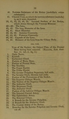
![40. Nervous Substance of the Retina [medullaris retina? substantia]. 41. Choroid Coat, on which the nervous substance [medulla] is, as it were, deposited. 39, 41, 23, 22, 46, 39. Internal Surface of the Retina, which shines through the Vitreous Humour. 42—46. The Lens. 43, 42. The long Diameter of the Lens. 44, 45. Short Diameter. 42, 44, 43. Anterior Convexity. 42, 45, 43. Posterior Convexity. 46, 26. Capsule of the Lens. 34, 42. Distance of the Lens from the Ciliary Body. FIG. VIII. View of the Socket; the Orbital Plate of the Frontal Bone having been removed. (Haller, Icon. Anat. fasc. vii. tab. vi. fig. 2.) 1. Optic Nerve. 2. Eyeball. 3. Lachrymal Gland. 4. Portion of Malar Bone. 5. Portion of Frontal Bone. 6. The Trochlea. 7. The Upper Eyelid. 8. The Under Eyelid. 9. The Levator Palpebrse Superioris, laid aside. 10. The Levator Oculi, likeudse laid aside. 11. Portion of the Periosteum of the Socket. 12. The Superior Oblique Muscle. 13. The Tendon of the Superior Oblique Muscle. 14. The Adductor Oculi. 15. The Depressor Oculi. 16. The Abductor Oculi. 17. Portion of the Inferior Oblique Muscle. 18. The Internal Carotid Artery. 19. The Ophthalmic Artery. 20. The External Ciliary. 21. A Branch for the Levator Palpebrse. 22. A Branch for the Abductor Oculi. 23. The Central Artery of the Retina. 24. Branches for the Levator Palpebrse and Levator Oculi, cut off.](https://iiif.wellcomecollection.org/image/b22386920_0040.jp2/full/800%2C/0/default.jpg)