Anatomical demonstrations, or, Colossal illustrations of human anatomy. Pt. I / by Professor Seerig ; translated from the German.
- Seerig, Albert Wilhelm Hermann, 1797?-1862.
- Date:
- 1831
Licence: Public Domain Mark
Credit: Anatomical demonstrations, or, Colossal illustrations of human anatomy. Pt. I / by Professor Seerig ; translated from the German. Source: Wellcome Collection.
Provider: This material has been provided by The Royal College of Surgeons of England. The original may be consulted at The Royal College of Surgeons of England.
40/40 (page 34)
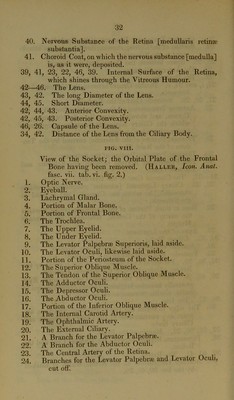
![8. A Branch, which goes into the hard Sheath [dura mater] of the Optic Nerve. 9. An Arterial Ring at the insertion of the Optic Nerve. 10, 11. The Long Ciliary Arteries. 12. Four Anterior Ciliaiy Arteries. 13. The Posterior Ciliary Arteries, which perforate the Sclerotic. FIG. X. The Cornea is removed, so as to show the Iris with its Vessels. (Haller, Icon. Anat. fasc. vii. tab. vi. fig. 6.) 1, 2. The Long Ciliary Arteries, which divide into two branches. 3. The Anterior Ciliary Arteries. 4. The Ciliary Ring. 5. The Larger Circle of the Iris. 6. A Portion of the Smaller Circle. [The whole circle cannot be seen, as all the vessels are not equally well filled.] 7. The Lens. J. mid C. Adlard, Printers, Bartholomew Close.](https://iiif.wellcomecollection.org/image/b22386920_0042.jp2/full/800%2C/0/default.jpg)