Licence: Public Domain Mark
Credit: A treatise on dislocations / by Lewis A. Stimson. Source: Wellcome Collection.
Provider: This material has been provided by the Royal College of Physicians of Edinburgh. The original may be consulted at the Royal College of Physicians of Edinburgh.
54/568
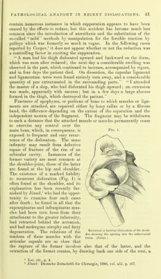
![The details of this formation, as observed by Baiardi' at the hip in animals, consist, first, in the appearance of a circular cartihiginous wall whose free border is continuous with the new-formed fibrous capsule, its base resting upon the ilium and its inner surface in contact with the head of the femur ; its ossification (in rabbits) is complete by the thirtieth day, except along its concave surface, where it remains soft, shading off toward the centre of the new acetabulum into a whitish, cartilaginous-like tissue, which takes the place of the destroyed periosteum. On its free border it has the structure of fibro-cartilage; on the concave surface it closely approximates that of hyaline, articular cartilage. At the very centre, as above said, the underlying bone is left bare or is covered by fibrous tissue and fibro-cartilage, and becomes denser in structure. Grinewetsky,^ who experimented on dogs, says he never found a lining of periosteum or cartilage inside the new acetabulum : the bone was always sclerosed. He also notes the absence of endothelium on the inner surface of the new capsule. The ossification may pass beyond the usual limits and include portions of the capsule,^ forming bony stalactites, or even a complete bony case enveloping, and perhaps united with, the head of the bone and in a specimen pi'esented by Moreau,^ a dislocation of the femur into the obturator foramen, the membrane filling the foramen had been trans- formed into a bony plate throughout, except in a strip along its anterior margin. Some of these experimental observations have been repeated upon specimens of ancient dislocations in man, in some of which the new cavity has been found to be lined with fibro-cartilage,^ in others with a granular fibroid tissue without apparent cartilage of incrustation.'^ The displaced head shows changes varying in extent and consisting in loss of its cartilage, erosion of the bone in places and its increase in others, and occasionally in profound changes of structure throughout. Thus in the case just i-eferred to, reported by Duguet, a dislocation inward of the shoulder of six months' standing, the head of the humerus was worn away behind at the point where it rested against the rim of the glenoid cavity, which also had in great part disappeared; its anterior portion had preserved its cartilage at almost all points, while its posterior portion had none, it being there replaced by rather tight, short fibrous bands uniting the head to the old glenoid cavity. In Lepine's case, a subcoracoid dislocation, the head of the humerus was considerably enlarged, Avith a deep vertical groove on its posterior surface corresponding to the outer edge of the new glenoid cavity, partly bare, partly covered by a fibrous layer. In a specimen presented by Walsh to the Royal Surgical Society of Ireland, 25th April, 1840,''' of an old dislocation of 1 Baiardi: Arch, [ler le Scienze niediche, 1880, vol. iv.; quoted by Kronlein. Grinewetsky : Centrnlblatt ftir Chirur<rie, 1879, p. 279. ' Thore: Bull, de la Sno. Anatoinique, ]'839, p. 33. ^ Cooper: Loc. cit., p. 50; and Cruveilliier : Auat. pathol., voL i. p. 425. 5 Moreau: Mgm. de I'Acad. royale de Chirurgie, 1769, vol, ii. p. 153. * Lepine and Desormeaux, in I?ull. de la Soc. Anat., 1844, p. 107. Duguet: Bull, de la Soc. Anat., 1803, p. 144. 8 Wiilsh: Gazette des Ilopit'iux, 1810, p. 330.](https://iiif.wellcomecollection.org/image/b21987063_0054.jp2/full/800%2C/0/default.jpg)