A peculiar form of hereditary congenital cataract / by E. Nettleship and F. Menteith Ogilvie.
- Edward Nettleship
- Date:
- [1906?]
Licence: In copyright
Credit: A peculiar form of hereditary congenital cataract / by E. Nettleship and F. Menteith Ogilvie. Source: Wellcome Collection.
Provider: This material has been provided by The Royal College of Surgeons of England. The original may be consulted at The Royal College of Surgeons of England.
13/24 page 5
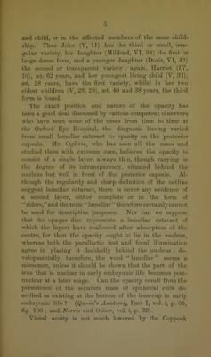
![and child, or in the affected members of the same child- ship. Thus John (V, 11) has the third or small, irre- gular variety, his daughter (Mildred, VI, 38) the first or large dense form, and a younger daughter (Doris, VI, 42) the second or transparent variety; again, Harriet (IV, 10), rnt. 62 years, and her youngest living child (V, 31), rnt. 28 years, have the first variety, whilst in her two eldest children (V, 26, 28), ast. 40 and 38 years, the third form is found. The exact position and nature of the opacity ha.s been a good deal discussed by various competent observers who have seen some of the cases from time to time at the Oxford Eye Hospital, the diagnosis having varied from small lamellar cataract to opacity on the posterior capsule. Mr. Ogilvie, who has seen all the cases and studied them with extreme care, believes the opacity to consist of a single layer, always thin, though varying in the degree of its intransparency, situated behind the nucleus but well in front of the posterior capsule. Al- though the regularity and sharp definition of the outline suggest lamellar cataract, there is never any evidence of a second layer, either complete or in the form of “riders,” and the term “lamellar” therefore certainly cannot be used for descriptive purposes. Nor can we suppose that the opaque disc represents a lamellar cataract of which the layers have coalesced after absorption of the centre, for then the opacity ought to lie in the nucleus, whereas both the parallactic test and focal illumination agree in placing it decidedly behind the nucleus ; de- velopmentally, therefore, the word “ lamellar ” seems a misnomer, unless it should be shown that the part of the lens that is nuclear in early embryonic life becomes post- unclear at a later stage. Can the opacity result from the ]iersistenco of the separate mass of epithelial cells de- scribed as existing at the bottom of the lens-cup in early embryonic life ? (J^unin’n Anatomy, Part I, vol. i, p. 85, fig. 100 ; and Norris and Oliver, vol. i, p. 39). Visual acuity is not mnch lowered by the Coppock](https://iiif.wellcomecollection.org/image/b22431202_0015.jp2/full/800%2C/0/default.jpg)

 No text description is available for this image
 No text description is available for this image
 No text description is available for this image