A system of human anatomy : general and special / by Erasmus Wilson.
- William James Erasmus Wilson
- Date:
- 1868
Licence: Public Domain Mark
Credit: A system of human anatomy : general and special / by Erasmus Wilson. Source: Wellcome Collection.
Provider: This material has been provided by the Francis A. Countway Library of Medicine, through the Medical Heritage Library. The original may be consulted at the Francis A. Countway Library of Medicine, Harvard Medical School.
50/660 (page 44)
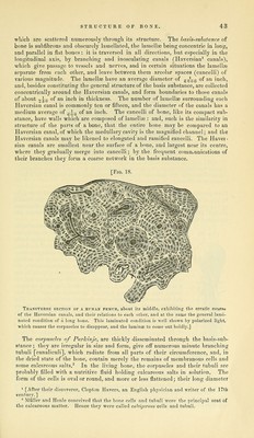
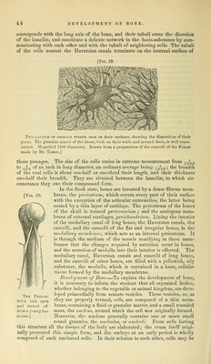
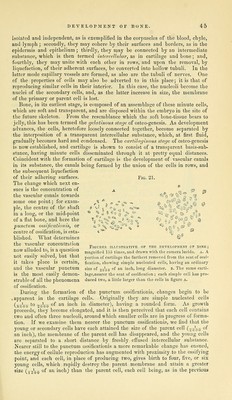
![corresponds with the long axis of the bone, and their tubuli cross the direction of the lamellte, and constitute a deHcate network in the basis-substance by com- municating with each other and with the tubuli of neighboring cells. The tubuli of the cells nearest the Haversian canals terminate on the internal surface of [Fig. 19. [Fig. 20. Two LACUNJE OF OSSEOUS TISSUE, seen on their surfaces, showing the disposition of their pores. The granular aspect of the tissue, both on their walls and around them, is well repre- sented. Magnified 1200 diameters. Drawn from a preparation of the cancelli of the Femur made by Mr. Tomes.] those passages. The size of the cells varies in extreme measurement from --q^qq to g-0y of an inch in long diameter, an ordinary average being jq^qq', the breadth of the oval cells is about one-half or one-third their length, and their thickness one-half their breadth. They are situated between the lamellae, to which cir- cumstance they owe their compressed form. In the fresh state, bones are invested by a dense fibrous mem- brane, the periosteum, which covers every part of their surface with the exception of the articular extremities, the latter being coated by a thin layer of cartilage. The periosteum of the bones of the skull is termed pericranium ; and the analogous mem- brane of external cartilages, pericliondrium. Lining the interior of the medullary canal of long bones, the Haversian canals, the cancelli, and the cancelli of the flat and irregular bones, is the rnxdullary membrane, which acts as an internal periosteum. It is through the medium of the vessels ramifying in these mem- branes that the changes required by nutrition occur in bones, and the secretion of medulla into their interior is effected. The medullary canal. Haversian canals and cancelli of long bones, and the cancelli of other bones, are filled with a yellowish, oily substance, the medulla, which is contained in a loose, cellular tis.«ue formed by the medullary membrane. Development of Bone.—To explain the development of bone, it is necessary to inform the student that all organized bodies, whether belonging to the vegetable or animal kingdom, are deve- loped primordially from minute vesicles. These vesicles, or, as they are properly termed, cells, are composed of a thin mem- brane, containing a fluid or granular matter, and a small rounded mass, the nucleus, around which the cell was originally formed. Moreover, the nucleus generally contains one or more small round granules, the nucleolus, or nucleoli. From cells having this structure all the tissues of the body are elaborated; the ovum itself origi- nally presented this simple form, and the embryo at an early period is wholly composed of such nucleated cells. In their relation to each other, cells may be Thk Perios- tultm 1 lid open and tu ^ned off from a y oung hu- merus.]](https://iiif.wellcomecollection.org/image/b21084452_0050.jp2/full/800%2C/0/default.jpg)