A system of human anatomy : general and special / by Erasmus Wilson.
- William James Erasmus Wilson
- Date:
- 1868
Licence: Public Domain Mark
Credit: A system of human anatomy : general and special / by Erasmus Wilson. Source: Wellcome Collection.
Provider: This material has been provided by the Francis A. Countway Library of Medicine, through the Medical Heritage Library. The original may be consulted at the Francis A. Countway Library of Medicine, Harvard Medical School.
53/660 (page 47)
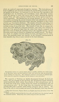
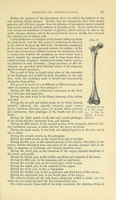
![[Fig. 24. During the progress of the phenomena above described, the contents of the cells undergo certain changes. At first, they are transparent, then they become granular, and still later opaque, from the presence of amorphous matter mingled with nuclei, nucleoli, and the remains of secondary colls. In the latter state the cells contain an abundance of minute oil-globules, which increase in size as the ossific changes advance, and in the newly-formed osseous areolae, have attained the ordinary size of adipose cells. Cartilaginification is complete in the human embryo at about the sixth week; and the first point of ossification is observed in the clavicle at about the fifth week. Ossification commences at the centre, and thence proceeds towards the surface; in flat bones the osseous tissue radiates between two membranes from a central point towards the periphery, in short bones from a centre towards the circumference, and in long bones from a central portion, diaphysis, towards a secondary centre, epiphy- sis, situated at each extremity. Large processes, as the tro- chanters, are provided with a distinct centre of development, which is named a'pophysis. The growth of bone in length takes place at the extremity of the diaphysis, and in bulk by fresh deposition on the sur- face; while the medullary canal is formed and increased by absorption from within. ^\i(iperiod of ossification is diiferent in different bones; the order of succession may be thus arranged : — During the fifth week, ossification commences in the clavi- cle, lower jaw, and upper jaw. During the sixth week, in the femur, humerus, tibia, radius, and ulna. During the seventh and eighth weeks, in the fibula, frontal, occipital, sphenoid, ribs, parietal, temporal, nasal, vomer, palate, vertebrae, first three pieces of sacrum, malar, metacar- pus, metatarsus, third phalanges of the hands and feet, and ilium. During the third month, in the first and second phalanges of the hands and feet, lachrymal bone, and ischium. During the fifth month, in the mastoid portion of the temporal, ethmoid, infe- rior turbinated, sternum, os pubis, and last two pieces of sacrum. During the sixth month, in the body and odontoid process of the axis, and in the OS calcis. During the seventh month, in the astragalus. During the tenth month, in the cuboid bone and os hyoides. During the first year, in the coracoid process of the scapula; first piece of the coccyx, inferior turbinated bone, last piece of the sternum, anterior arch of the fttlas, OS magnum, os unciforme, and external cuneiform bone. During the third year, in the cuneiform of the carpus, internal cuneiform of the tarsus, and patella. During the fourth year, in the middle cuneiform and scaphoid of the tarsus, During the fifth year, in the trapezium and os semilunare. During the seventh year, in the second piece of the coccyx. During the eighth year, in the scaphoid of the carpus. During the ninth year, in the os trapezoides. During the twelfth year, in the os pisiforme and third piece of the coccyx. During the eighteenth year, in the fourth piece of the coccyx. The ossicula auditus are the only bones completely ossified at birth ; the ver- tebrae are not completed until the five-and-twentieth year. The entire osseous framework of the body constitutes the skeleton, which, m A YOUNG Femur. 1, 5. The epiphy- ses. 4. The din- physis. 2, 3. Apo- physes.]](https://iiif.wellcomecollection.org/image/b21084452_0053.jp2/full/800%2C/0/default.jpg)