The fundus oculi with an ophthalmoscopic atlas, illustrating its physiological & pathological conditions / By W. Adams Frost.
- Frost, W. Adams (William Adams), 1853-1935
- Date:
- 1896
Licence: Public Domain Mark
Credit: The fundus oculi with an ophthalmoscopic atlas, illustrating its physiological & pathological conditions / By W. Adams Frost. Source: Wellcome Collection.
Provider: This material has been provided by The University of Leeds Library. The original may be consulted at The University of Leeds Library.
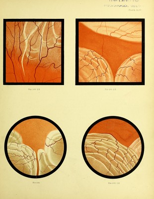
429/448
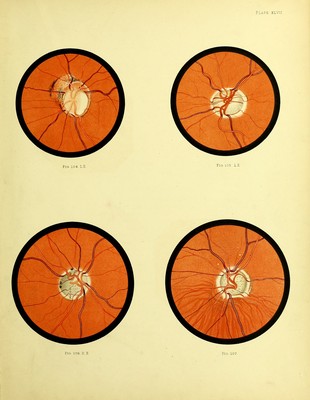
![COLOBOMA OF DISC. GLAUCOMA SIMPLEX. Fig. 104.—Partial Colo bo ma of Disc. Mary S., 15. LE. Drawing made August 1889.—A healthy looking girl. No signs of inherited syphilis, and no likelihood of the disease having been acquired. The RE. had been defective as long as the patient can remember. There were large areas of choroidal atrophy. The physiological cup on the disc appeared to be filled up with a material which could not be well defined with the ophthalmoscope. The LE. (that depicted) V. = f. Visual field normal. Description.—The disc is of unusually large size. The greater part of it is deeply cupped. The uncupped portion forms a crescent widest above; its limbs pass round the disc forming a ring which becomes very narrow on the lower and outer border. The vessels curve abruptly over the well defined edge of the cup. The floor of the cup presents on its temporal side a white crescent. On the upper and inner side of, and in contact with, the disc, is a patch of partial atrophy of the choroid. Fig. 105.—Glaucomatous Cup. Alfred H., 41. LE. Dnming made 1888.—In the winter of 1879-80, after exposure to bad weather, he became suddenly blind whilst driving. There was no pain, and the sight returned in about three hours. In the spring of 1882, after much anxiety, the sight of the left eye gradually failed, the failure being accompanied by halos round a light. In the winter of 1883-4 there was gradual failure in both eyes with pain in the eyes and head. Mr. Streatfeild performed iridectomy on the LE., and a few months later on the BE. The operations were followed by considerable improvement, so that he could read and write. Vision has, howevei', gradually failed since. Descriptiun.—Disc oval, surrounded by a halo of choroidal atrophy. The whole disc is cupped, the larger vessels bending abruptly over its edge. The floor of the cup has a some- what greenish tint, and is stippled. Fig. 106.—Glaucomatous Cup. Thomas S., 46. BE. Drawing made October 1888.—First seen December 1887. Vision failing gradually two or three years, more rapidly the last year. No pain in the eyes, but occasional frontal pain. Both sclei'otics were of a bluish grey colour. Although the patient did not look older than his years, there was a complete arcus senilis in both eyes. This formed a sharply defined ring, about a millimetre wide, and extending right up to the sclero-corneal junction. BE. = 4r] T + 2. Inner half of visual field lost (see Chart). L^E. +2-25 D = ^^; T.n. Visual field normal. The ophthalmoscopic appearance in did](https://iiif.wellcomecollection.org/image/b21512656_0429.jp2/full/800%2C/0/default.jpg)