Demonstrations of anatomy : being a guide to the dissection of the human body / by G.V. Ellis.
- George Viner Ellis
- Date:
- 1887
Licence: Public Domain Mark
Credit: Demonstrations of anatomy : being a guide to the dissection of the human body / by G.V. Ellis. Source: Wellcome Collection.
Provider: This material has been provided by The University of Leeds Library. The original may be consulted at The University of Leeds Library.
125/790 (page 111)
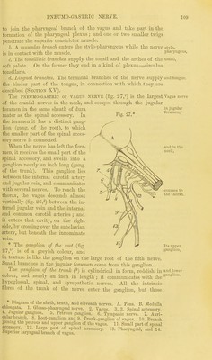
![the sympathetic; and lower down it supplies twigs to the inferior constrictor. c. Cardiac branches. One or two small cardiac nerves spring Brandies to from the pneumo-gastric at the upper part of the neck, and join {^p^J^ cardiac branches of the sympathetic. At the lower part of the neck, lower. on each side, there is a larger cardiac nerve which descends into the thorax :—the right one joins the deep nerves to the heart from the sympathetic; and the left terminates in the superficial cardiac plexus. d. The inferior or recurrent laryngeal nerve leaves the pneumo-gastric Lower trunk on the right side opposite the subclavian artery, and winding J^^1 to round that vessel, takes an upward course in the neck to the larynx, ascending beneath the common carotid and inferior thyroid arteries, along the groove between the trachea and the oesophagus. At the larynx it enters beneath the ala of the thyroid cartilage, where it will be afterwards traced. The following branches arise from it :— Some' cardiac branches leave the nerve as it turns round the sub- gives clavian artery ; these enter the thorax, and join the cardiac nerves heart 6810 of the sympathetic. Tracheal and oesophageal branches spring from it as it ascends in the to trachea, neck ; and near the larynx some filaments are furnished to the ^ p1]^8' inferior constrictor muscle. rynx. On the left side the recurrent nerve arises in the thorax, opposite Left reeur- the arch of the aorta ; in the neck it lies between the trachea and r nerve, oesophagus, as on the right side. The SPINAL ACCESSORY NERVE Courses through the jugular Eleventh foramen with the pneumo-gastric, but is not marked by any ganglion. nerve The nerve is composed of two parts, viz., a smaller one, accessory to has two the vagus, and a larger, spinal part, which have a different origin p and distribution. The part accessory to the vagus (fig. 27,u) arises from the medulla Accessory oblongata, and ends by joining the pneumo-gastric outside the skull. to vagus In the foramen of exit it lies close to the vagus, and is connected to in foramen: the upper ganglion of that nerve by one or two filaments. Below beIow the foramen it passes over the lower ganglion of the vagus, and ' 'en' blends with the trunk beyond that ganglion. It gives distinct off- sets to join the pharyngeal and upper laryngeal branches of the pneumo-gastric ; and other fibres are continued into the cardiac and inferior laryngeal branches. The spinal part (fig. 27,12), which takes its origin from the spinal Spinal part cord, is much larger, and is connected with the smaller piece while m foramen: passing through the jugular foramen. Beyond the foramen the in the neck nerve (fig. 26,2) takes a backward course through the sterno-mastoid, and across the side of the neck to end in the trapezius : at first it is crosses to concealed by the jugular vein, but it then passes either over or under trapc2IUS' that vessel. The connections of the nerve beyond the sterno- mastoid have been already examined (p. 56). The nerve furnishes muscular offsets to the sterno-mastoid and supplies the trapezius. muscles. The hypoglossal nerve, issuing from the cranium by the Twelfth](https://iiif.wellcomecollection.org/image/b21518439_0125.jp2/full/800%2C/0/default.jpg)