Demonstrations of anatomy : being a guide to the dissection of the human body / by G.V. Ellis.
- George Viner Ellis
- Date:
- 1887
Licence: Public Domain Mark
Credit: Demonstrations of anatomy : being a guide to the dissection of the human body / by G.V. Ellis. Source: Wellcome Collection.
Provider: This material has been provided by The University of Leeds Library. The original may be consulted at The University of Leeds Library.
138/790 (page 124)
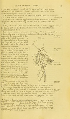
![Upper con- strictor arises from pterygoid process, jaw and tongue. Inserted partly into a raphe. Relations. Interval between muscle and skull. Use of constrictor: in swallow- ing of constrictor. Dissection to show longitudinal muscles. between the origins of the middle and inferior constrictors are the j superior laryngeal nerve and vessels. The superior constrictor is thinner than the others, and of a ] quadrilateral form. It has a broad origin from the following parts in succession, commencing above,—the lower end of the internal pterygoid plate and the hamular process, the pterygo-maxillary ligament, the hinder part of the mylo-hyoid ridge of the lower jaw, the mucous membrane of the mouth, and the side of the tongue. The fibres pass backwards, and are inserted by joining those of the fellow muscle along the middle bine, where a tendinous raphe is formed between the two for the upper half of their depth. Some of the highest fibres end on the aponeiu'osis of the pharynx. The parts in contact with this muscle externally are the deep vessels and nerves of the neck at the side, the middle constrictor and prevertebral muscles behind : internally are the aponeurosis of the pharynx and the palato-pharyngeus muscle. The upper border forms an arch with the concavity upwards extending from the pterygoid plate to the basilar process ; and the space between it and the base of the skull is occupied by the aponeurosis of the pharynx, which projects outwards above the muscle, and by the levator palati, Eustachian tube and inferior palatine artery. The attachment to the pterygo-maxillary ligament corresponds with the origin of the buccinator muscle (i). Action of constrictors. The muscles of both sides contracting at' the same time will diminish the size of the pharynx ; and as the anterior attachments of the lower muscles are nearer together than those of the upper, the tube will be contracted more behind the larynx than near the head. In swallowing, the object is first seized by the lower part of the upper constrictor, and then forced on to the oesophagus by the sue-' cessive action of the middle and inferior constrictors. Since the back of the pharynx is closely applied to the prevertebral muscles, from which it cannot be separated in the natural condition of the parts, the effect of the contraction of these muscles is to draw the tongue, hyoid bone and larynx backwards, as well as somewhat upwards owing to the oblique direction of the greater number of the fibres of the middle and lower constrictors ; and the cavity, when empty, is compressed from before backwards. The upper part of the superior constrictor narrows the space- above the mouth, and assists in bringing together the posterior pillars of the soft palate. (See the action of the palato-pharyn- geus.) Dissectim. By dividing the middle and inferior constrictor* midway between their origin and insertion, and reflecting the parts forwards and backwards, the longitudinal fibres of the pharyngeal wall will be exposed. The longitudinal or elevator muscles of the pharynx are the stylo-pharyngeus and palato-pharyngeus. The styloyharyngcus has already been described, but it may now be followed to its insertion (p. 102). The palofo-pharyngevs is only partially seen, and will be](https://iiif.wellcomecollection.org/image/b21518439_0138.jp2/full/800%2C/0/default.jpg)