Demonstrations of anatomy : being a guide to the dissection of the human body / by G.V. Ellis.
- George Viner Ellis
- Date:
- 1887
Licence: Public Domain Mark
Credit: Demonstrations of anatomy : being a guide to the dissection of the human body / by G.V. Ellis. Source: Wellcome Collection.
Provider: This material has been provided by The University of Leeds Library. The original may be consulted at The University of Leeds Library.
33/790 (page 19)
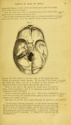
![The NINTH Or GLOSSOPHARYNGEAL, the TENTH, PNEUMO-GASTRIC Ninth, or VAGUS, and the ELEVENTH or SPINAL ACCESSORY NERVES (fig. 4, 8) eleventh*1 pass through the middle compartment of the jugular foramen. The {^ou^-h aS* glosso-pharyngeal is external to the other two, and has a distinct jugular opening in the dura mater. The spinal accessory nerve ascends foramen- through the foramen magnum and, together with the vagus, enters an aperture in the dura mater close to the occipital bone. The TWELFTH or HYPOGLOSSAL NERVE (fig. 4, 9) is the motor Twelfth nerve of the tongue, and consists of two small pieces, which pierce neive- the dura mater separately opposite the anterior condylar foramen ; these unite at the outer part of that aperture. Dissection. The dissector may now return to the examination of Dissection the trunk of the carotid artery as it winds through the cavernous 0 carotld- sinus. On the opposite side of the head, on which the nerves in the ofsympa- wall of the cavernous sinus are untouched, an attempt may be made uses'? plex to find two small plexuses of the sympathetic on the carotid artery, though in an injected body this dissection is scarcely possible. One of these (cavernous) is near the root of the anterior clinoid cavernous, process ; and to bring it into view it will be necessary to cut off that piece of bone, and to dissect out with care the third, fourth, fifth, and sixth nerves, looking for filaments between them and the plexus. Another plexus (carotid), joining the fifth and sixth and carotid, nerves, surrounds the artery as it enters the sinus. The internal carotid artery appears in the cranium at the J11^'™1 apex of the petrous part of the temporal bone. In this part of its artery course the vessel lies between the layers of the dura mater bound- ing the cavernous sinus along the side of the body of the sphenoid bone, and makes two bends so as to have the form of the letter S reclined. It first ascends in the inner part of the foramen lacerum, winds and then runs forwards to the root of the anterior clinoid process ; cavernous finally it turns upwards in the groove on the inner side of this pro-sinus- cess, perforates the dura mater forming the roof of the sinus, and divides into cerebral arteries at the base of the brain. In this course the artery is enveloped by nerves derived from the sympa- thetic in the neck (p. 114). The branches of the artery here are few. In the sinus there are Branches, some small arteries (arterise receptaculi) for the supply of the dura mater and the bone, the nerves, and, the pituitary body ; and oppo- site the anterior clinoid process the ophthalmic branch arises. The terminal branches of the carotid will be seen in the dissec- tion of the base of the brain (p. 184). Sympathetic Nerve. Accompanying the carotid arteiy is a pro- Sympathetic iongation of the sympathetic nerve of the neck, which 'forms the forms following plexuses :— The carotid plexus is situate on the outer side of the vessel, at ™rotM its entrance into the cavernous sinus, and communicates with the P 6XUS' sixth nerve and the Gasserian ganglion. The small cavernous plexus is placed below the bend of the artery cavernous which is close to the anterior clinoid process, and is mainly derived ]>ll'iUS; o 2](https://iiif.wellcomecollection.org/image/b21518439_0033.jp2/full/800%2C/0/default.jpg)