A Text-book of medical practice for practitioners and students / edited by William Bain.
- Date:
- 1904
Licence: In copyright
Credit: A Text-book of medical practice for practitioners and students / edited by William Bain. Source: Wellcome Collection.
Provider: This material has been provided by The University of Leeds Library. The original may be consulted at The University of Leeds Library.
40/1042 page 12
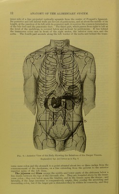
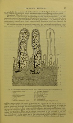
![inner side (if a line projected vertically upwards from the centre of Poupart's ligament. Its posterior and left lateral walls are devoid of peritoneum, and at about the middle of its length, at the junction of its left with its posterior wall, it receives the common termination of the bile duct and the pancreatic duct. The tliird part, which passes from right to left at the level of the umbilicus, is covered below and in front l)y peritoneum. It lies behind the transverse colon and in front of the right ureter, the inferior vena cava and the aorta. The fourth part ascends along the left border of the aorta and behind the trans- ■piQ_ 9. Anterior View of the Body Showing the Relations of the Deeper Viscera. Explanatory figs, and letters as in Fig. 6. verse meso-colon and the stomach to a point situated about two or three inches from the commencement of the duodenum, on a line extemling from the pylorus tt) the anterior superior s])inc of the left ilium. , The jejuniim and ilium occupy the middle and lower parts of the abdomen below a line drawn'l)etwcen the tips of the eleventh ribs. They are bounded above by the trans- verse colon ; they rest l)elow upon the bladder, and in the female upon the utei-us ; anrt laterally, if the large intestine is comparatively empty, they overlai) the a.scenduig and descending colon, but if the larger gut is distended they lie witlnn its concavity, an.i tliey](https://iiif.wellcomecollection.org/image/b21510167_0040.jp2/full/800%2C/0/default.jpg)