A Text-book of medical practice for practitioners and students / edited by William Bain.
- Date:
- 1904
Licence: In copyright
Credit: A Text-book of medical practice for practitioners and students / edited by William Bain. Source: Wellcome Collection.
Provider: This material has been provided by The University of Leeds Library. The original may be consulted at The University of Leeds Library.
44/1042 page 16
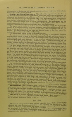
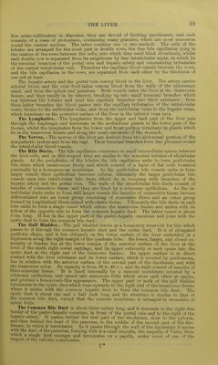
![it is embraced by the internal and external sphincters, between which some of the anterior fibres of the levator ani are inserted. Structure and (Jenenil Appearance.—The walls of the large intestine like those of the stomach and small intestine consist of four chief coats, the serous, the muscular, the submucous and the mucous, but unlike the small intestine its walls are sacculated and the sacculi are arranged in three longitudinal rows, which are separated from each other by three Imnds of longitudinal muscle fibres. In the rectum the sacculations are not in rows and the longitudinal bands unite to form a continuous coat. The anal passage and the A'ermiform appendix are devoid of sacculi and have the longitudinal fibres of their external muscular coats arranged in a continuous layer, as in the small intestine. The serous coat is similar to that on the small intestine, except that it gives off a number of processes filled with fat which are called appendices epiploicfe, and it is de- ficient on the posterior aspects of the ascending colon, the descending colon and the rectum. It is also absent from the sides and anterior surface of the lower part of the rectum and from the walls of the anal passage. The submucosa is similar to that of the small intestine. The mucous membrane is smooth, devoid of villi and pitted by the orifices of numerous tubular glands, similar to those of the small intestine, but longer and more numerous, and containing many more mucous cells. It contains many solitary follicle.s similar to those met with in the small intestine. In the vermiform appendix these follicles are so numerous that they practically form a continuous layer. After the thirtieth year the solitary follicles undergo atrophy and in old age they are less numerous and less pro- minent than in middle life. The surface of the membrane is covered with columnar cells. The septa which intervene between the sacculations of the large intestine consist, mainly, of folds of the mucous and submucous coats, with some of the circular fibres of the muscular coat, and although the rows of sacculations disappear in the rectum the cavity of this part of the large intestine is usually separated into four sections, of which the lowest and largest is called the ampulla, by three semilunar folds, the valves of Houston, which are similar in structure to the septa between the sacculations of the colon. The middle and largest of these three folds is situated on the right side of the rectum at the level of the reflection of the peritoneum from the rectum to the bladder or to the vagina, and the two smaller folds are on the left side one above and one below the large fold on the right side. These folds are of importance inasmuch as the end of any instrument intr(xluced into the rectum may catch against them and tearing through their bases enter the extra-peritoneal tissue or even the peritoneal cavity. The vascular sui)]jly of the large intestine is important, more especially in association with the anastomosis which takes place round the lower part of the rectum and m the walls of the anal passage between the tributary veins of the portal and systemic systems and with the fact that the veins of the portal system are devoid of valves. Ilius it is that in any obstruction to the blood flow through the liver the anastomoses between the superior hi^morrhoidal veins and the inferior hsemorrhoidal veins become dilated, as the blood which should have passed through the liver to the inferior vena cava is f^.rced through these relatively small passages towards the internal pudic and iliac veins, it the distention of the anastomosing passages is l..ng continued piles are gradually formed. At the same time it should be noted that if the obstruction to the portal cn:culation is the liver the superficial veins round the umbilicus will also be dilated by blood which has passed to them through the anastomosing channels which run along tli« r^d h^^^ ment from the left branci of the portal vein (see below), whilst if the »b«^ruction is outside the liver, and afi'ects the trunk of the portal vein, the superficial veins aiound the umbilicus will not be implicated. _ . i.„ ;„ fu^ rrlanrl« The Lyn.phatics.-The lymphatics of the vermiform appendix terminate m the gkn^^ in the lowest part of the mesentery. The lymph vessels of the f ^^^^^^^.^fj^^f'T^^^^ lie along the posterior aspect of this portion of the alimenfciry canal ^^^^/^ ^^f J^^^/^^^^^^^^ efferent vesse s issue which terminate in the lumbar glands. The Ij'^Ph * passes through glands which lie in the concavity of the sacrum to the pre-^ortic glands, and the lymphatics of the anal passage terminate m the pubic groups of ^^^^^^^^^^^^^^^ The Nerves of the large intestine are derived from the ^^^^'^.^^^^^ji^^^^^^^^ plexuses and from the second, third and fourth sacral nerves. Their arrangement in the wall of the large intestine is similar to that met with m the small intestine. The Livbe, The liver is a large mass of very vascular glandular tissue. It lies im^^^^^^ epigastric and right hypochondriac Sa^ in tL^IS^^^ extent into tlie right lumbar region, and its left extremity extends, rcguiaiiy and occasionally in the adult, into the left hypochondrium. neritoneum and it Except on the right part of its posterior surface it is is attached to the difphragm, the back of tlie Imea alba and the stomach by tokls pen](https://iiif.wellcomecollection.org/image/b21510167_0044.jp2/full/800%2C/0/default.jpg)