A text-book on surgery : general, operative, and mechanical / by John A. Wyeth.
- John Allan Wyeth
- Date:
- 1898
Licence: Public Domain Mark
Credit: A text-book on surgery : general, operative, and mechanical / by John A. Wyeth. Source: Wellcome Collection.
Provider: This material has been provided by the Francis A. Countway Library of Medicine, through the Medical Heritage Library. The original may be consulted at the Francis A. Countway Library of Medicine, Harvard Medical School.
21/1024
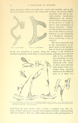
![modic contraction of the terminal arterioles, the capillaries, and vennles in the injured area, followed almost instantly by dilatation of these ves- sels far beyond their normal caliber. The volume of blood is at once greatly increased to fill the enlarged channels, and the current is more rapid, since capillary resistance is less. After the lapse of about one hour the current begins to slacken and gradually becomes slower than before the injury. This slowing of the current is not due to recon- traction of the vessels, but to a clogging of their channels with the corpuscular elements of the blood. The red blood-corpuscles and the plaques * floating in the plasma occupy the center of the vessels, while the white corpuscles (leuco- cytes), which normally exist in the blood in the proportion of from 1 to 1,000 to 1 to 250 of the red corpuscles, are largely increased and are seen to adhere to the ves- sel walls (Fig. 3). Since the force of the current is greatest in the arterioles and least in the venules (which have the capillaries between them and the heart to retard the cir- culation), the leucocytes can not adhere to the lining membrane of the arterioles ; a consider- able number are seen attached to the capil- laries, while the venules are practically choked with them, and it is through the walls of the venules that they emigrate (diapedesis, Stair?]- Bdv, to ooze through) and wander into the in- tervascular spaces (Fig. 3.) Some few pass through the capillaries, but none have been observed to escape through the walls of the arterioles. The leucocyte, which has the power of changing its form (Fig. 10), pushes through the line of union of the flat epithelium which composes the wall of the venule, displacing the cement substance, and finally emerging at the outer side of the vessel, which by its elastici- ty at once closes the aperture of escape. Co- incident with the clogging of the venules and the emigration of the leucocytes, by reason of the force of the heart's action, the plasma oozes Fig. 3.—Inflamed mesentery of a frog. F, vein; A, small artery and capillaries. The red corpuscles are seen in the center of the current; the white blood-corpuscles creep along their inner walls, some being in the process of emigration; the surround- ing tissues contain many of these which have al- ready emigrated from the vessels. (Tillmann.) Fig. 4.—Diapedesis or emigration of leucocytes through the walls of a venule, a, incomplete, b, com- plete emigration (schematic). (Tillmann.) * The plaques or third corpuscles of the blood measure from 1-3 to 3-5 micromillimetres in diameter, and are supposed to be embryonic red blood-corpuscles. They consist of a colorless pro-](https://iiif.wellcomecollection.org/image/b21085213_0021.jp2/full/800%2C/0/default.jpg)