Rest and pain : a course of lectures on the influence of mechanical and physiological rest in the treatment of accidents and surgical diseases, and the diagnostic value of pain / by the late John Hilton ; edited by W.H.A. Jacobson.
- John Hilton
- Date:
- 1896
Licence: Public Domain Mark
Credit: Rest and pain : a course of lectures on the influence of mechanical and physiological rest in the treatment of accidents and surgical diseases, and the diagnostic value of pain / by the late John Hilton ; edited by W.H.A. Jacobson. Source: Wellcome Collection.
60/538 page 40
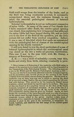
![fluid could cf^cape from the interior of the brain; and as the fluid was being constantly secreted, it necessarily accumulated there, and the occlusion formed, to my mind, the essential pathological element of internal hydrocephalus. Internal hydrocephalus is not an unfrequent companion of spina bifida. In many of the cases of that disease that I have examined after death, the cerebro-spinal opening was closed, thus explaining how it happened that although the spina bifida had been tapped during life, and no limit ]uit to the flow of the fluid at the operation, yet the little patient did not suffer from cerebral congestion. Indeed, it was a case of this kind which first made me acquainted with the pathological fact of closure of the cerebro-spinal opening in the fourth ventricle.* I will now detail to you the short particulars of a case of internal hydrocephalus in which the cerebro-spinal canal was closed at the iter a tertio ad quartum ventriculum. This case I saw in the year 1847, with Mr. Otway, a surgeon at Kennington:— A. H. was a child of unhealthy parents, very thin, feeble and sickly from birth, uttering constantly a pecu- * Other instanf'ps of distension of the ventricles with fluid owing tp closure of the aperture in tlie fourth ventricle, will be found recorded in the 1'ransactions of the Patliological Society. The following cases are g ven in Dr, Wilk’s and Dr. Moxon’s Pathological Anatomy, p. 215 :— “ In one case the brain was firmly adherent round the foramen magnnm, and the membranes and choroid plexus at the opening of the fourth ventricle were very thick, almost closing the aperture. But the most re- markable case we have seen was one in which on opening the skull tin re appeared a large bladder like an hydatid separating the left cere- bral convolutions. This proved to be the immensely dilated lateral veutricle—which had thrust its way through to the surface. The cause of this was found to be a papillose tumour in the floor of the fourth ventiicle, composed of ependymal structure. It was of the size of a small hazel-nut, and completely cut off the cavity of the ventricle. Two remarkably similar cases are given by Virchow. Again, we have known the ventricles of the brain distended much in a case of pressure on the spinal canal through disease of the cervical spine, thus cerebral symptoms became added to the spinal. Another certain cause of expansion of the ventricles is tumour or tubercle under the tentorium ; whether it grow in the pons or the cerebellum, it compresses the fourth ventricle, and disallows the free escape of the cerebro-spinal fluid.”—[Ed.]](https://iiif.wellcomecollection.org/image/b28136718_0060.jp2/full/800%2C/0/default.jpg)


 No text description is available for this image
 No text description is available for this image
 No text description is available for this image