An American text-book of surgery : for practitioners and students / By Phineas S. Conner, M.D., Frederic S. Dennis, M.D., William W. Keen, M.D., Charles B. Nancrede, M.D., Roswell Park, M.D., Lewis S. Pilcher, M.D., Nicholas Senn, M.D., Francis J. Shepherd, M.D., Lewis A. Stimson, M.D., J. Collins Warren, M.D., and J. William White, M.D. Ed. by William W. Keen and J. William White.
- William Williams Keen
- Date:
- 1899
Licence: Public Domain Mark
Credit: An American text-book of surgery : for practitioners and students / By Phineas S. Conner, M.D., Frederic S. Dennis, M.D., William W. Keen, M.D., Charles B. Nancrede, M.D., Roswell Park, M.D., Lewis S. Pilcher, M.D., Nicholas Senn, M.D., Francis J. Shepherd, M.D., Lewis A. Stimson, M.D., J. Collins Warren, M.D., and J. William White, M.D. Ed. by William W. Keen and J. William White. Source: Wellcome Collection.
Provider: This material has been provided by the Augustus C. Long Health Sciences Library at Columbia University and Columbia University Libraries/Information Services, through the Medical Heritage Library. The original may be consulted at the the Augustus C. Long Health Sciences Library at Columbia University and Columbia University.
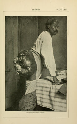
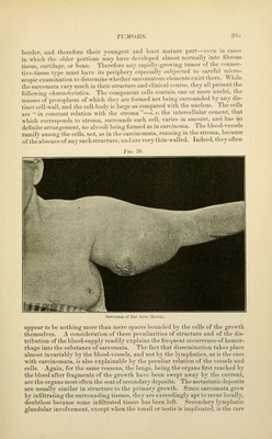
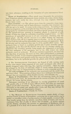
![border, and therefore their youngest and least mature part—even in cases in wliieii the ohler portions may have developed almost normally into fibrous tissue, cartilage, or bone. Therefore any rapidly-growing tumor of the connec- tive-tissue tyi)e nmst have its periphery especially subjected to careful micro- scopic examination to determine whether sarcomatous elements exist there. AVhde the sarcomata vary much in their structure and clinical course, they all ])resent the following characteriiitics. The component cells contain one or more nuclei, the masses oi' protoplasm of which they are formed not being surrounded by any dis- tinct cell-wall, and the cell-body is 'large as compared with the nucleus. The cells are '^ in constant relation with the stroma—/, e. the intercellular cement, that which corresi)oruls to stroma, surrounds each cell, varies in amount, and has no definite arrangement, no alveoli being formed as in carcinoma. The blood-vessels ramify among the cells, not, as in the carcinomata, running in the stroma, because of the absence of any such structure, and are very thin-walled. Indeed, they often Fig. 29. .■sarcoma of the Arm ^Keun). appear to be nothing more than mere spaces bounded by the cells of the growth themselves. A consideration of these peculiarities of structure and of the dis- tribution of the blood-supply readily explains the frequent occurrence of hemor- rhage into the substance of sarcomata. The fact that dissemination takes place almost invariably by the blood-vessels, and not by the lymphatics, as is the case with carcinomata, is also explainable by the peculiar relation of the vessels and cells. Again, for the same reasons, the lungs, being the organs first reached by the blood after fragments of the growth have been swept away by the current, are the organs most often the seat of secondary deposits. The metastatic deposits are usually similar in structure to the primary growth. Since sarcomata grow by infiltrating the surrounding tissues, they are exceedingly apt to recur locally, doubtless because some infiltrated tissue has been left. Secondary lymphatic glandular involvement, except when the tonsil or testis is implicated, is the rare](https://iiif.wellcomecollection.org/image/b21217014_0261.jp2/full/800%2C/0/default.jpg)