An American text-book of surgery : for practitioners and students / By Phineas S. Conner, M.D., Frederic S. Dennis, M.D., William W. Keen, M.D., Charles B. Nancrede, M.D., Roswell Park, M.D., Lewis S. Pilcher, M.D., Nicholas Senn, M.D., Francis J. Shepherd, M.D., Lewis A. Stimson, M.D., J. Collins Warren, M.D., and J. William White, M.D. Ed. by William W. Keen and J. William White.
- William Williams Keen
- Date:
- 1899
Licence: Public Domain Mark
Credit: An American text-book of surgery : for practitioners and students / By Phineas S. Conner, M.D., Frederic S. Dennis, M.D., William W. Keen, M.D., Charles B. Nancrede, M.D., Roswell Park, M.D., Lewis S. Pilcher, M.D., Nicholas Senn, M.D., Francis J. Shepherd, M.D., Lewis A. Stimson, M.D., J. Collins Warren, M.D., and J. William White, M.D. Ed. by William W. Keen and J. William White. Source: Wellcome Collection.
Provider: This material has been provided by the Augustus C. Long Health Sciences Library at Columbia University and Columbia University Libraries/Information Services, through the Medical Heritage Library. The original may be consulted at the the Augustus C. Long Health Sciences Library at Columbia University and Columbia University.
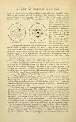
![a much deeper and more irregular coloring than is usually seen in acute inflammation. Redness is entirely absent in bloodless parts, as in the cor- nea, but in this case we find a hyperemia of the vessels of the conjunctiva, and later an actual development of vascular loops in the direction of the inflamed spot. It was at one time su])posed that the increased warmth of the part was due to a local production of heat. It is now known that the local rise of temperature is due to the greater amount of blood which flows throu'^h the vessels. One of the most constant symptoms of inflammation is the swelling. This is rarely absent, and is seen even in non-vascular parts. The increase<l amount of blood in the vessels of the part does not add materially to its size. We must seek for an explanation of this phenomenon in the altered condition of the tissues of the part. On making an incision into an inflamed spot we find the meshes of tissue distended with an abundant exudation of blood-serum and leucocytes escaping through the walls of the dilated blood-vessels. The tissues are saturated with this material often to such an extent that it may be difticult for the surgeon to recognize the difference between muscles, fasciie, and vessels. The exudation consists not only of leucocytes, but, in addition, of a certain amount of fluid which closely resembles the liquor sanguinis, and from which fibrin is formed giving a certain firmness to the part. The tissues are also crowded with leucocytes. The increased number of cells in the part was at one time attributed to the division or proliferation of the pre-existing cells of tlie inflamed tissues, but Cohnheim maintained tliat the new cells were the escaped Avhite blood-corpuscles, and that the so-called fixed connective-tissue cells played no part in the process, being incapable of proliferation. This doctrine he illus- trated by experiments upon the cornea. The opacity produced by an artificial inflammation was found to be due to the presence of numberless leucocytes, while the corneal corpuscles were found to be unchanged. Subsequent obser- vations have, however, shown that the fixed cells of the cornea also undergo proliferation and take part in the process. Cohnheim thought that the immense number of cells found in an inflamed part were all derived from the white cor- puscles, and that by subsequent proliferation they were increased in number and formed what is known as granulation-tissue^ which he assumed played a prominent part in the healing process. It is a well-known fact, however, at the present time, that the fixed connective-tissue corpuscles and other cells in tissues of the body are capable of division. According to the latest views on the origin of the granulation-tissue, the round cells with single nuclei are mostly formed by the proliferation of connective-tissue and other fixed-tissue cells. Later, many of these cells, as also the leucocytes, become polynuclear cells, and as such are incapable of taking any further active part in the process. Accord- ing to Ziegler, the polynuclear leucocytes appear to be taken up and destroyed by the proliferating connective-tissue cells, the leucocytes apparently serv- ing simply as nutriment for these cells. The process of multiplication by cell-division is now much better understood than formerly, and the mode of indirect division (karyokinesis, p. 29) in which the nucleus plays a prominent part is the one most frequently observed. The meshes of the tissue are distended with coagulated lymph, and the connective-tissue fibres are swollen and softer than usual, and here and there terminate suddenly, as if broken off, giving them a club-shaped appearance. In the organs aflected we find the epithelial cells altered in appearance, being in the condition known as that of cloudy swelling; that is, their protoplasm is granular and more opaque, and containing frequently fatty granules. Dur- ing the development of the inflammatory process the leucocytes are seen infil-](https://iiif.wellcomecollection.org/image/b21217014_0047.jp2/full/800%2C/0/default.jpg)