A treatise on the blood, inflammation, and gun-shot wounds / by the late John Hunter. To which is prefixed, A short account of the author's life, by his brother-in-law, Everard Home.
- John Hunter
- Date:
- 1794
Licence: Public Domain Mark
Credit: A treatise on the blood, inflammation, and gun-shot wounds / by the late John Hunter. To which is prefixed, A short account of the author's life, by his brother-in-law, Everard Home. Source: Wellcome Collection.
Provider: This material has been provided by University of Bristol Library. The original may be consulted at University of Bristol Library.
651/674 page 569
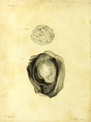
![Z 569 ] [PLATE THIRD. This represents a front view of the human testicle, upon the body of which is a coagulum of blood adhering to it. For the better under- standing this plate it will be necessary to give at length the history of the case. A man came into St. George's Hospital with an hydrocele, for which he was tapped with a lancet. When the water was evacuated, the tes- ticle was larger to the feel than common, and in a month the tunica va- ginalis was as much distended as before the operation. The radical cure was now determined upon ; the tunica vaginalis was slit open, but the testicle being enlarged it was thought proper to extract it. Upon the body of the testicle was found a coagulum of blood, resembling a leech in appearance, and in the angle between the testicle and epididymis, was another smaller one ■ at some parts it adhered to the testicle and epidi- dymis and at others it was loose from both. The adhesion of the large coagulum was firm, although it admitted of a separation, which was made at one end; when separated, fibres were plainly seen running between it and the testicle. The adhesions of the small coagulum were in marry places still firmer. This blood had been extra- vasated by the puncture made with the lancet in drawing off the water, and had fallen down upon the testicle, where it coagulated. Over the whole surface of the tunica vaginalis there were vessels filled with blood, and clots of extravasated blood in different parts. figure r. The testicle, with the tunica vaginalis slit open, exposing its surface. AA. The body of the testicle. B. A small hydatid arising from its surface, which occurs not unfre- quently in that situation, viz. just where the epididymis takes its origin from the testicle. C. The smaller coagulum lying in the angle between the body of the testicle and the epididymis. 4D](https://iiif.wellcomecollection.org/image/b2144111x_0651.jp2/full/800%2C/0/default.jpg)


 No text description is available for this image
 No text description is available for this image
 No text description is available for this image