Text-book of ophthalmology / by Ernst Fuchs ; authorized translation from the eleventh revised and greatly enlarged German edition with numerous additions by Alexander Duane.
- Ernst Fuchs
- Date:
- [1911]
Licence: In copyright
Credit: Text-book of ophthalmology / by Ernst Fuchs ; authorized translation from the eleventh revised and greatly enlarged German edition with numerous additions by Alexander Duane. Source: Wellcome Collection.
Provider: This material has been provided by UCL Library Services. The original may be consulted at UCL (University College London)
989/1032 page 953
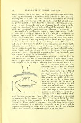
![The operation of Panas tries to secure the connection between the lid and the frontalis muscle by the formation of a pedicle from the skin of the former, which pedicle is attached to the skin of the forehead and to the surface of the muscle. Fig. 441 shows how the pedicle is cut out of the skin of the lid. The pedicle, s, after being defined by incision, is dissected from its bed until it is freely movable; then a horizontal incision, a, is made through the skin directly above the eyebrow. Start- ing both from this incision and from the wound already made below, the skin of the eyebrow is undermined so that a bridge of tissue is formed, beneath which the pedicle, s, is slipped so that its upper margin is in contact with the upper lip of the incision, a. Its attachment to the latter is effected by means of a loop of thread, the center of which lies on the cutaneous side of the pedicle, while its extremities, b b, are passed through the upper lip of the wound. By drawing the loop tight the pedicle is lifted up and is attached to the upper border of the wound. If necessary, a second loop may be applied, and also some interrupted sutures, to secure exact adaptation of the edges. The operation produces a satisfactory effect but has the disad- vantage of leaving scars which run perpendicular to the direction of the fibers of the orbicularis and hence are pretty conspicuous. 3. The superior rectus is also available for replacing the levator. Following Motais's method, we first expose this muscle by cutting through the conjunctiva at a point corresponding to the insertion of the tendon, and starting from this incision we make a second incision in the con- junctiva along the muscle itself carrying it back through the upper retrotarsal fold and as far as the convex border of the tarsus. The tendon is now detached from the sclera along a line occupying the middle of its insertion and 3 to 4 mm. wide; and by starting from each extremity of this incision and splitting the muscle longitudinally far backward, we fashion out of its middle third a free tongue, while the nasal and tem- poral thirds of the muscle remain connected with the sclera. The free end of the tongue is then stitched to the upper border and the anterior surface of the tarsus. [According to the somewhat different description given by Bruns, this is done by passing a double-armed thread through the tendon near its main insertion and in that part which forms the free tongue. (The latter may be cut either before or after passing the stitch, and should extend back as far as possible.) The thread is firmly tied, and the ends are left long. Then with a blunt instrument the skin is separated from the tarsus down to the free border of the lid. In the space thus opened up, the two needles are carried, brought out just above the free border of the lid, and tied, over a roll of gauze tight enough to produce a marked primary over-effect. The conjunctival wound, especially in the cul-de-sac, must be carefully closed.—D.]](https://iiif.wellcomecollection.org/image/b21287454_0991.jp2/full/800%2C/0/default.jpg)
 No text description is available for this image
 No text description is available for this image
 No text description is available for this image