Contributions to the physiology and pathology of the breast and its lymphatic glands / by Charles Creighton.
- Charles Creighton
- Date:
- 1886
Licence: Public Domain Mark
Credit: Contributions to the physiology and pathology of the breast and its lymphatic glands / by Charles Creighton. Source: Wellcome Collection.
163/262 (page 143)
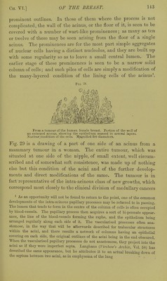
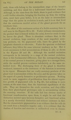
![prominent outlines. In those of them where the process is not complicated, the wall of the acinus, or the floor of it, is seen to be covered with a number of wart-like prominences ; as many as ten or twelve of these may be seen arising from the floor of a single acinus. The prominences are for the most part simple aggregates of nuclear cells having a distinct nucleolus, and they are built up with some regularity so as to leave a small central lumen. The earlier stage of these prominences is seen to be a narrow solid column of cells; and such piles of cells are simply a modification of the many-layered condition of the lining cells of the acinus\ Fig. 29. From n liimour of tlie human female breast. Portion of thn wmII of aJi enlarged acnms, showing the epithelium massed in several layers. ]Vuclear_coudition of the cells. Magnified 300 diameters. Fig. 29 is a drawing of a part of one side of an acinus from a mammary tumour in a woman. The entire tumour, which was situated at one side of the nipple, of small extent, well circum- scribed and of somewhat soft consistence, was made up of nothing else but this condition of the acini and of the further develop- ments and direct modifications of the same. The tumour is in fact representative of the intra-acinous class of new growths, which correspond most closely to the clinical division of medullary cancers 1 As an opportunity will not be found to return to the point, one of the common developments of the intra-acinous papillary processes may be referred to in passing. The lumen that tends to form in the centre of the column of cells is often occupied by blood-vessels. The papillary process then acquires a sort of bi-pennate appear- auce, the hne of the blood-vessels forming the raphe, and the epithehum being arranged regularly along each side of it. The vascularised processes often ana- stomose, in the way that will be afterwards described for trabecular structm-es within the acini, and there results a network of columns having an epithehal covering on each side, the original outlines of the acini bemg in the end obscm-ed. When the vascularised papillary processes do not anastomose, they project into the acini as if they were imperfect septa. Langhans (Virchow's Archiv, Vol. 58) haa described the same appearance, but he attributes it to an actual breaking down of the septiun between two acini, as in emphysema of the lung](https://iiif.wellcomecollection.org/image/b20415321_0163.jp2/full/800%2C/0/default.jpg)