Licence: Public Domain Mark
Credit: The anatomy of the human body / By J. Cruveilhier. Source: Wellcome Collection.
Provider: This material has been provided by the University of Massachusetts Medical School, Lamar Soutter Library, through the Medical Heritage Library. The original may be consulted at the Lamar Soutter Library at the University of Massachusetts Medical School.
112/944 (page 88)
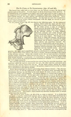
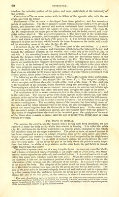
![The Os Coxce, or Os Innominatum {figs. 47 and 48). The liawnch hone, called also os coxcb, from coxa, the haunch, occupies the lateral and anterior parts of the pelvis. It is the largest of all the broad bones of the skeleton. It is asymmetrical, very irregular in its figure, and twisted upon itself, so that it appears to be composed of two portions ; one superior, triangular, shaped like a wing, and flattened from without inward; the other inferior, and flattened from before backward. These two parts are united by a contracted portion. On this bone we have for consideration an external or femoral surface, which corresponds with the thigh, an internal or pelvic surface, and a circumference. 0 1 tl e femoral surface {fig 47) we observe the following parts : On the contracted portion, which unites the upper and the lower half of the os coxae, is the cotyloid cavity (a, figs. 47, 48) (from kotvTit], a cup), or acetabulum, which receives the head of the femur. This is of a hemispherical shape, and is the deepest of all the articular cavities ; it looks obliquely downward, outward, and a little forward, and has a considerable depression (b, fig. 47) at the bottom, on its inner aspect. The margin (c) of this cavity is sharp and sinuous, and presents three notches, or, rather, one notch and two slight depressions, one of which is superior, and the other inferior, and somewhat external. The notch (d) is situated below and on the inside ; it is very deep, and converted into a foramen by a ligament, and gives passage to the vessels which penetrate into the cotyloid cavity. Above and below the acetabulum are two horizontal grooves , the upper one is superficial and gives attachment to a fibrous expansion, one of the origins of the rectus femoris; the lower is deeper, and gives passage to the tendon of the obturator externus. Above the cotyloid cavity, the os coxze presents a broad tri- angular surface, improperly called external iliac fossa (e). On it we observe, proceeding from behind forward, 1. A convexity : 2. A concavity, occupying about two thirds of the fossa, and on which the principal nutritious foramina are situated : 3. A second convex- ity : 4. A slight concavity. The external iliac fossa is traversed by two curved lines for muscular insertions ; one posterior, called the superior semicircular liiie (/); the other anterior, and much more extensive, the inferior semicircular line (g). All the whole surface behind the former gives attachment to the gluteus maximus : the portion comprised between the two lines gives attachment to the gluteus medius. Below the acetabulum we observe proceeding from without inward, 1. The obturator foramen {h, figs. 47 and 48), improperly so called ; it is placed more internally than the acetabulum, and has an oval form in the male (hence its name, foramen ovale): in the female it is smaller, and triangular. Its longest diameter is vertical, and it slopes a lit- tle downward and outward. At its upper part is the obturator groove {i, fig. 48), directed obliquely from behind forward and inward. It gives passage to vessels and nerves, and has two lips: an anterior, which is continuous with the outer half of the circumference of the foramen ; and a posterior, which is continuous with the internal half; for the two halves of the circumference of the obturator foramen, instead of being united in front, pass in difierent directions, the internal backward, and the external forward, leaving be- tween them an interval which constitutes the groove. 2. On the inside of the obturator foramen is a square surface {k,figs. 47 and 48), broader above than below, oblong in a vertical direction, and rough for the insertion of several muscles of the thigh. This is continuous below with another surface (l, fig. 47), broader inferiorly than above, which extends obliquely downward and outward, then curves upward, and terminates below the cotyloid cavity. This surface, which bounds the obturator foramen below, is intend- ed for muscular insertions. The internal or pelvic surface (fig. 48) of the os coxae is concave. It is divided into two parts : a superior, which looks upward, and an inferior, which looks backward, by a prominent horizontal ridge (m n. o, fig. 48), which forms the lower boundary of the in- ternal iliac fossa. Above this ridge, which we shall afterward see forms the greatest part of the inlet of the pelvis, we find, proceeding from behind forward, 1. A very prom- inent and rough tuberosity (r), intended for several ligamentous attachments. 2. An ir- regular articular surface, broader above than below, and called the auricular surface (s) of the OS coxae, from a supposed resemblance to the concha of the ear; it looks down- ward and inward, and articulates with a corresponding surface on the sacrum. 3. More anteriorly, and on a higher plane, a deep and regular excavation, correctly denominated the internal iliac fossa {t). This fossa, which is broad and smooth, is occupied by the](https://iiif.wellcomecollection.org/image/b21196801_0112.jp2/full/800%2C/0/default.jpg)