Licence: Public Domain Mark
Credit: The anatomy of the human body / By J. Cruveilhier. Source: Wellcome Collection.
Provider: This material has been provided by the University of Massachusetts Medical School, Lamar Soutter Library, through the Medical Heritage Library. The original may be consulted at the Lamar Soutter Library at the University of Massachusetts Medical School.
187/944 (page 163)
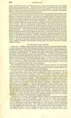
![Fig. 78, part of the corfesponding cavity uncovered.* In this respect ihe inter-articular cartilages of the knee differ from all others of the same kind, for they do not establish a complete separ- ation of the articular surfaces, between which they are placed. These falciform cartilages are inserted into the tibia by means of ligaments, which deserve a particular description. Ligaments ef the External Semilunar Cartilage.—These are two : the one anterior, and the other posterior ; both of them are very strong. The anterior is inserted in front of the spine of the tibia, outside of the anterior crucial ligament, into a deep depression situated near the external glenoid cavity of the tibia. This anterior ligament of the external semilunar cartilage sends off a bundle which intermingles with the ante- rior crucial ligament. Tlie posterior is inserted into the spine of the tibia, in the unequally-divided interval situated between the two prominences of the spine. The posterior ligament sends off a considerable bundle of fibres to be inserted into the posterior crucial ligament. The circular form of the external semilunar cartilage is owing to the insertions of the two an- terior and posterior ligaments being separated from each other only by a few lines. Ligaments of the Internal Semilunar Cartilage.—These are much weaker than the for- mer. The anterior is inserted a good deal before its fellow, the anterior ligament of the external semilunar cartilage, and the posterior is inserted a good deal behind the corresponding ligament of the external semilunar cartilage ; hence the semilunar shape ■of the internal semilunar cartilage, which does not send off any fibrous prolongation to the anterior or posterior crucial ligaments. The ligaments of the inter-articular cartila- ges being inserted into the tibia, these cartilages follow the tibia throughout its course. Means of Union of the Kfiee-joint are two lateral ligaments, a posterior and an anterior, two crucial ligaments, and a synovial capsule. 1. Lateral Ligaments.—The external lateral ligament {a, Jigs. 79 and 80) appears as a rounded cord; it is inserted into the exter- nal tuberosity of the femur, at the point of union of the five anterior sixths with the first posterior, on the prolongation of the line of the fibula ; the precise point of this insertion is a small eminence surmounting a depression which is destined to the tendon of the popli- teus muscle, and is situated in front of an- other depression destined to the external ge- mellus ; thence this ligament descends, in a vertical line, to be inserted mto the external face of the head of the fibula. This ligament has the appearance of a tendon; it extends along the anterior border of the tendon of the biceps, with which it may be readily con- founded. We should have but an incomplete idea of the means of union which the knee-joint pos- sesses on the outside, if we did not add to the number of its ligaments the tendon of the bi- ceps, which unites in some sort its inferior insertions with those of the external lateral ligament, and the small band of the fascia lata inserted into the anterior tubercle of the tibia^ and sending to the external edge of the rotula an expansion, which unites with the tendon of the vastus externus The internal lateral ligament (b c, figs. 79 and 80), which is much longer than the exter- nal, has the shape of a broad, thin, pearly- coloured band, arising from the posterior part Fig. 79. Fig. 80. * On asking myself the question why there should be this difference between the two semilunar cartila<;es, I have come to the conclusion that the external condyle of the femur, pressing much more upon the tibia than the intemai, on account of the external following the axis of the femur, while the internal is turned away from it to the inside, the external iater-artictilar cartilage had to piotect a greater ] the tibia rtion of the articular surface of](https://iiif.wellcomecollection.org/image/b21196801_0187.jp2/full/800%2C/0/default.jpg)