Licence: Public Domain Mark
Credit: The anatomy of the human body / By J. Cruveilhier. Source: Wellcome Collection.
Provider: This material has been provided by the University of Massachusetts Medical School, Lamar Soutter Library, through the Medical Heritage Library. The original may be consulted at the Lamar Soutter Library at the University of Massachusetts Medical School.
222/944 (page 198)
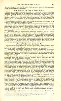
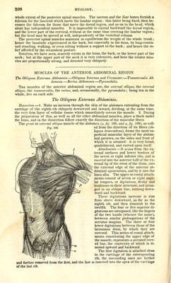
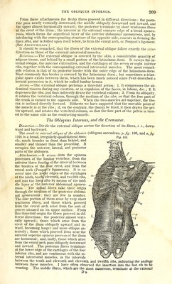
![MUSCLES OF THE POSTERIOR REGION OF THE TRUNK. The Trapezius.—Latissimus Dorsi and Teres Majm.—Rhomboideus.—Levator Anguli ScapulcB. Serrati Postici.—Splenius.—Posterior Spinal Muscles.—Complexus.—Inter- spinalis Colli.—Recti Capitis Postici, Major et Minor.—Ohliqui Capitis, Major et Minor. General View and Action of the Posterior Spinal Muscles. The muscles situated on the posterior region of the trunk form several layers, which, proceeding from the skin to the bones, consist, on either side, of the trapezius, the latis- simus dorsi and teres major, the rhomboideus and levator anguli scapulae, the serrati postici, superior and inferior, the splenius, the long muscles of the back, viz., the sacro- lumbal'is and longissimus dorsi; the transversalis colli and the complexus (which I re- gard as two series of accessory fasciculi to the longissimus dorsi) ; the complexus ma- jor, the inter-spinales coUi, the recti capitis postici, major et minor, and the obliqui capitis, major et minor.* The Trapezius. Dissection. 1. Render the muscle tense by placing a block under the chest; 2. Make an incision through the skin from the occipital protuberance to the twelfth dorsal verte- bra and another horizontally from the seventh cervical vertebra to the external end of the'clavicle ; 3. Reflect the two flaps, together with the cellular membrane adhering in- timately to the muscle ; 4. Dissect very carefully the insertions into the occipiteil bone, which consist of a very thin aponeurosis closely united to the skin. The trapezius (cucuUaris, Albinus, a, Jigs. 106,113), the most superficial muscle on the •* -,^„*«-;^— «^™;,,.« «r*u« 4'».-.»i* rt^.tr^-.-i> Fig. 106. posterior region of the trunk, covers the nape of the neck and the back. It is a broad triangular, rather than trapezoid muscle, thick in the mid- dle, thin and elongated at its supe- rior and inferior angles. Attachments.—It arises from the spinous processes of all the dorsal and the seventh cervical vertebrae, from the corresponding supra-spi- nous ligaments, from the posterior cervical ligament (ligamentum nu- chcE;, and from the internal third of the superior occipital line, and is in- serted into the entire length of the spine of the scapula, into the poste- rior border of the acromion, and into the external third of the posterior border of the clavicle. The fixed attachments or origins of this mus- cle present, 1. A broad, semi-ellipti- cal aponeurosis, which, when united to the one on the opposite side, forms an ellipse, occupying the space be- tween the sixth cervical and the third dorsal vertebrae ; 2. A very thin fibrous lamina, not having the ordi- nary shining appearance of an apo- neurosis, which is firmly adherent to the skin, and forms the truncated occipital angle of the muscle ; 3. A great number of tendmous fibres, constituting all those attachments to the vertebra; that are independ- ent of the two preceding aponeuro- ses. From these origins all the fleshy fibres proceed mitward, the mferior fibres from below upward, the superior from above downward, and from behind forward, and the middle ones horizontally. They terminate in the following manner : the lower or ascending fibres are collected together, and at- tached to a triangular aponeurosis, which, gliding over the small facette at the internal extremity of the spine of the scapula, is inserted into the tubercle immediately connect- ed with it; the middle or horizontal fibres terminate at the posterior border of the spine of the scapula, by tendinous fibres which are very distinct, especially towards the acro- * [The transversa-spinalis muscle includes the semi-spinalis colli, the semi-spinalis dorsi, and the multifidas spinse of Alhinus.]](https://iiif.wellcomecollection.org/image/b21196801_0222.jp2/full/800%2C/0/default.jpg)