Licence: Public Domain Mark
Credit: The anatomy of the human body / By J. Cruveilhier. Source: Wellcome Collection.
Provider: This material has been provided by the University of Massachusetts Medical School, Lamar Soutter Library, through the Medical Heritage Library. The original may be consulted at the Lamar Soutter Library at the University of Massachusetts Medical School.
237/944 (page 213)
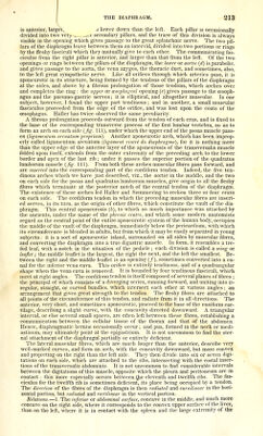
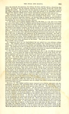
![\ is anterior, largeY, d lower down than the left. Each pillar is occasionally- divided into two verj--, .oc secondary pillars, and the trace of this division is always visible in the opening which gives passage to the great splanchnic nerve. The two pil- lars of the diaphragm leave between them an interval, divided into two portions or rings by the fleshy fasciculi which they mutually give to each other. The communicating fas- ciculus from the right pillar is anterior, and larger than that from the left. Of the two openings or rings between the pillars of the diaphragm, the lower or aortic (d) is parabolic, and gives passage to the aorta, the vena azygos, the thoracic duct, and sometimes, also, to the left great sympathetic nerve. Like all orifices through which arteries pass, it is aponeurotic in its structure, being formed by the tendons of the pillars of the diaphragm at the sides, and above by a fibrous prolongation of those tendons, which arches over and completes the ring : the upper or cesophag-cal opening (e) gives passage to the oesoph- agus and the pneumo-gastric nerves; it is elliptical, and altogether muscular. In one subject, however, I found the upper part tendinous; and in another, a small muscular fasciculus proceeded from the edge of the orifice, and was lost upon the coats of the oesophagus. Haller has twice observed the same peculiarity. A fibrous prolongation proceeds outward from the tendon of each crus, and is fixed to the base of the corresponding transverse process of the first lumbar vertebra, so as to form an arch on each side (fig. Ill), under which the upper end of the psoas muscle pass- es {ligam-entum arcnatum proprium). Another aponeurotic arch, which has been improp- erly called ligamentum arcuatum {ligament cintri du diaphragme), for it is nothing more than the upper edge of the anterior layer of the aponeurosis of the transversa] is muscle folded upon itself, extends from the outer extremity of the preceding arch to the lower border and apex of the last rib ; under it passes the superior portion of the quadratus lumborum muscle {fig. 111). From both these arches muscular fibres pass forward, and are inserted into the corresponding part of the cordifonn tendon. Indeed, the five ten- dinous arches which we have just described, viz., the aortic in the middle, and the two on each side for the psoas and quadratus lumborum muscles, give origin to all the fleshy fibres which terminate at the posterior notch of the central tendon of the diaphragm. The existence of these arches led Haller and Soemmering to reckon three or four crura on each side. The cordiform tendon in which the preceding muscular fibres are insert- ed serves, in its turn, as the origin of other fibres, which constitute the vault of the dia- phragm. This central aponeurosis (J), to which so much importance was attached by the ancients, under the name of the phrenic centre, and which some modern anatomists regard as the central point of the entire aponeurotic system of the human body, occupies the middle of the vault of the diaphragm, immediately below the pericardium, with which its circumference is blended in adults, but from which it may be easily separated in young subjects : it is a sort of aponeurotic island, surrounded on all sides by muscular fibres, and converting the diaphragm into a true digastric muscle. In form, it resembles a tre- foil leaf, with a notch in the situation of the pedicle ; each division is called a wing or leaflet; the middle leaflet is the largest, the right the next, and the left the smallest. Be- tween the right and the middle leaflet is an opening (/), sometimes converted into a ca- nal for the inferior vena cava. This orifice is entirely tendinous, and of a quadrangular shape when the vena cava is removed. It is bounded by four tendinous fasciculi, which meet at right angles. The cordiform tendon is itself composed of several planes of fibres ; the principal of which consists of a diverging series, running forward, and uniting into ir- regular, straight, or curved bundles, which intersect each other at various angles ; an arrangement that gives great strength to the tendon. The fleshy fibres are attached to all points of the circumference of this tendon, and radiate from it in all directions. The anterior, very short, and sometimes aponeurotic, proceed to the base of the ensiform car- tilage, describing a slight curve, with the concavity directed downward. A triangular interval, or else several small spaces, are often left between these fibres, establishing a communication between the cellular tissue of the thorax and that of the abdomen. Hence, diaphragmatic herniae occasionally occur ; and pus, formed in the neck or medi- astinum, may ultimately point at the epigastrium. It is not uncommon to find the ster- nal attachment of the diaphragm partially or entirely deficient. The lateral muscular fibres, which are much longer than the anterior, describe very well-marked curves, and form an arch, with the concavity downward, but more convex and projecting on the right than the left side. They then divide into six or seven digi- tations on each side, which are attached to the ribs, intersecting with the costal inser- tions of the transversalis abdominis. It is not uncommon to find considerable intervals between the digitations of this muscle, opposite which the pleura and peritoneum are in contact: this more especially occurs between ^he eleventh and twelfth ribs. The fas- ciculus for the twelfth rib is sometimes deficient, its place being occupied by a tendon. The direction of the fibres of the diaphragm is then radiated and curmlinear in the hori- zontal portion, but radiated and rectilinear in the vertical portion. Relations.—1. The inferior or abdominal surface, concave in the middle, and much more concave on the right side, where it corresponds to the convex upper surface of the liver, than on the left, where it is in contact with the spleen and the large extremity of the](https://iiif.wellcomecollection.org/image/b21196801_0237.jp2/full/800%2C/0/default.jpg)