Licence: Public Domain Mark
Credit: The anatomy of the human body / By J. Cruveilhier. Source: Wellcome Collection.
Provider: This material has been provided by the University of Massachusetts Medical School, Lamar Soutter Library, through the Medical Heritage Library. The original may be consulted at the Lamar Soutter Library at the University of Massachusetts Medical School.
292/944 (page 268)
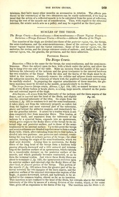
![fleshy fasciculi, accessories to the obturator intemus, are generally distinguished by anat- omists into the superior (/) and the inferior (g); they are separated from each other by the tendon of the obturator intemus, for the reception of which they form a groove. Above and below this groove they take their origin ; the superior from the spine of the ischium, and the inferior, which is the larger, from the tuberosity of that bone, immedi- ately above the attachment of the great sacro-sciatic ligament, and even slightly from the ligament itself. They both pass horizontally outward, are sometimes united either behind or in front of the tendon of the obturator intemus, which they then completely embrace, and with which they are entirely or partially blended, being inserted with it into the digital cavity of the great trochanter. Their relations are the same as those of the reflected portion of the obturator intemus. The gemellus superior is often wanting, and the inferior is frequently double. I have several times seen the superior terminate in the tendon of the pyriformis, and the in- ferior in the tendon of the obturator intemus. Action.—They rotate the thigh outw-ard. Their relations with the synovial capsule of the obturator intemus led to their being designated marsupiales by Cotoper, and by Portal, le muscle capsulaire de la capsule du tendon de I'obturateur interne. The Quadratus Femoris. This muscle (i, fig. 125), shaped like a parallelogram, is situated immediately below the gemellus inferior. It arises from the extemal border of the tuberosity of the ischium, in front of the semi-membranosus, from which it is separated by adipose tissue. From this point the fibres proceed horizontally outward, parallel to each other, and are inserted into an oblong ridge* projecting partly from the back of the root of the great trochan- ter, and partly from the femur inmiediately below it; but above the attachment of the adductor magnus, with which, at first, it appears to be continuous, but from which it is always separated by the internal circumflex vessels. This muscle is sometimes wanting ; but very frequently its pelvic attachments are pro- longed as far as the ascending ramus of the ischium ; in which cases it is twisted inferi- orly upon itself, so as to oppose a surface, not a border, to the adductor magnus. Its re- lations behind are the same as those of the preceding muscles ; in front, it covers the obturator externus and the lesser trochanter, from which it is often separated by a sy- novial capsule. The Obturator Externus. Dissection.—The lower or horizontal portion of the obturator externus is exposed, by dividing the quadratus femoris into two equal parts by a vertical incision. In order to see the upper or pelvic portion, it is necessary to detach the gracilis, pectineus, psoas, Ui- acus, and adductor brevis. This is a triangular, flat muscle (e, fig. 127), of the same shape, but thinner and smaller than the obturator internus, and, like it, reflected, though at an obtuse angle. It arises from the circumference of the obturator foramen, from the obturator ligament, and from the tendinous arch which completes the sub-pubic canal for the vessels and nerve. It is inserted into the deepest and lowest part of the digital cavity of the great trochanter. The fleshy fibres arise directly, the lower ones proceed horizontally outward, and the upper obliquely downward, backward, and outward; thus converging, they form a fleshy belly, which turns round the neck of the femur, and terminates in a tendon that passes horizontally outward, to be inserted into the digital cavity, below the gemelli and the ob- turator internus. Relations.—Its outer and anterior surface is in relation with the pectineus, the adduc- tors, the psoas and iliacus muscles, and more externally with the neck of the femur and the lower part of the capsular ligament of the hip-joint. Its inner and posterior surface is in contact with the obturator foramen and the quadratus muscle. Action of the preceding Muscles. The last six muscles are evidently rotators of the thigh outward. The pyriformis, the gemelli, and the obturator internus, which are almost always united at their insertions, would deserve the name of quadri-gemini, given by the older anatomists to the gemeUi, the pyriformis, and the quadratus. WTien they take their fixed point upon the femur, as, for example, in standing upon one foot, they become rotators of the pelvis, and turn the anterior surface of the trunk to the opposite side. They are only rotators when the limb is extended ; in the sitting posture, they become abductors. Winslow, who first demon- strated their use in abduction in the semiflexed position, attached great importance to the connexion of so many of these muscles with the capsular ligament, which he believed prevented pinching of the capsule during the different movements of the joint. The insertion of these muscles is exceedingly favourable. Moreover, we shall find, that besides the glutaeus maximus and the posterior fibres of the glutaeus medius and * [M. Cruveilhier states the insertion of the quadratus femoris to be into the inter-trochanteric line. The description in the text, copied from Albinus, gives a more accurate idea of the insertion of this muscle.]](https://iiif.wellcomecollection.org/image/b21196801_0292.jp2/full/800%2C/0/default.jpg)