Licence: Public Domain Mark
Credit: The anatomy of the human body / By J. Cruveilhier. Source: Wellcome Collection.
Provider: This material has been provided by the University of Massachusetts Medical School, Lamar Soutter Library, through the Medical Heritage Library. The original may be consulted at the Lamar Soutter Library at the University of Massachusetts Medical School.
67/944 (page 43)
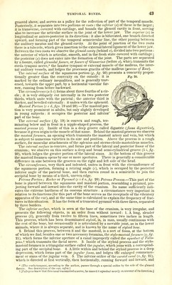
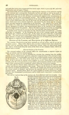
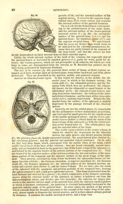
![grooved above,' and serves as a pulley for the reflection of part of the temporal muscle. Posteriorly, it separates into two portions or roots: the inferior (o) of these is the larger; it is transverse, covered with cartilage, and bounds the glenoid cavity in front, serving also to increase the articular surface in the joint of the lower jaw. The superior («) is longitudinal or antero-posterior in its direction : it also is bifurcated, one branch directed upward, and forming part of the temporal semicircular Une, the other passing between the auditory meatus and the glenoid cavity. At the point of junction of the two roots there is a tubercle, which gives insertion to the external lateral ligament of the lower jaw. Between the two roots we observe the glenoid cavity (behind o), divided into two portions : the anterior of which is articular, smooth, and in the fresh state covered with cartilage ; the posterior (s) does not enter into the formation of the joint. The parts are separated by a fissure, called glenoidal fissure, or fissure of Glasserms (before s), whicli transmits the corda tympani nerve,* the laxator tympani or external muscle of the malleus, the inter- nal auditory vessels, and lodges the processus gracilis of the malleus {p-ocess of Raw). The internal surface of the squamous portion {g, fig. 20) presents a concavity propor- tionally greater than the convexity on the outside: it is marked by the ordinary inequalities, and is generally trav- ersed, towards the upper part, by a horizontal vascular fur- row, running from before backward. The circumference (a h c) forms about three fourths of a cir- / cle; it is very obliquely cut internally in its two posterior , thirds, which unite with the parietal; the anterior third is ' thicker, and bevelled externally: it unites with the sphenoid. I Mastoid Portion (c e d,figs. 19 and 20).—The mastoid por- Sj^, tion is very prominent in adults, but only slightly developed in young subjects : it occupies the posterior and inferior' part of the bone. The external surface {fig. 19) is convex and rough, ter- minating below and in front in a nipple-shaped process, the mastoid process (e). Inside of this is a deep groove called digastric {fossa digasfrica), because it gives origin to the muscle of that name. Behind the mastoid process we observe the mastoid foramen, an opening which transmits the mastoid artery and vein, but which is subject to numerous varieties in its size and position. Above the process is a rough surface, for muscular attachments of the splenius and sterno-cleido mastoideus muscles. The internal surface is concave, and forms part of the lateral and posterior fossaj of the cranium ; we observe on this surface a deep and broad semi-cylindrical groove {h i, fig. 20), which lodges the greater portion of the lateral sinus. At the bottom of this groove the mastoid foramen opens by one or more apertures. There is generally a considerable difference in size between the grooves on the right and left side of the head. The circumference, very thick and indented, unites in front with the circumference of the squamous portion, forming a retiring angle (c), which is occupied by the posterior inferior angle of the parietal bone, and then curves round in a semicircle to join the occipital bone by means of a thick, uneven edge. Petrous Portion; Rochcr or Pyramid (c i d v, fig. 20) Petrous Process.—This part of t'ne bone is placed between the squamous and mastoid portion, resembling a pyramid, pro- jecting forward and inward into the cavity of the cranium. Its name sufficiently indi- cates the extreme hardness of its osseous structure : a circumstance very important in relation to its functions (for this part of the bone serves as the receptacle of the vibratory apparatus of the ear), and at the same time is calculated to explain the frequency of frac- tures in this situation. It has the form of a truncated pyramid with three faces, separated by three borders. The inferior surface, which is seen at the base of the cranium, is very irregular, and presents the following objects, in an order from without inward: 1. A long, slender process {k), generally from twelve to fifteen lines, sometimes two inches in length. This process, which has been denominated styloid, is, in man, usually continuous with the rest of the bone, but occasionally it is articulated by a movable joint, as in the lower animals, where it is always separate, and is known by the name of styloid bone. 2. Behind this process, between it and the mastoid, is a sort of fossa, at the bottom of which we find, besides one or two accessory foramina, the stylo-mastoid foramen {y. fig. 21), which forms the inferior aperture of a canal improperly called the aqueduct of Fallo- pius,i which transmits the facial nerve. 3. Inside of the styloid process and the stylo- mastoid foramen is a triangular surface called the jugular, which joins with a correspond- ing part of the occipital bone. 4. A little within and behind the styloid process is a deep depression, which forms part of the jugular fossa, and lodges tKe enlarged commence- ment or sinus of the jugular vein. 5. The inferior orifice of the carotid canal {v,fig. 21), which is directed at first vertically, then horizontally, running forward and inward, and * [The corda tympani, according to the author, passes through a special orifice by the side of the glenoid fissure. See description of the ear, tn/rd.] 1 [Fullopius knew that this canal transmitted a nerve; he named it aqueduct merely on account of its direction.]](https://iiif.wellcomecollection.org/image/b21196801_0067.jp2/full/800%2C/0/default.jpg)