Licence: Public Domain Mark
Credit: The anatomy of the human body / By J. Cruveilhier. Source: Wellcome Collection.
Provider: This material has been provided by the University of Massachusetts Medical School, Lamar Soutter Library, through the Medical Heritage Library. The original may be consulted at the Lamar Soutter Library at the University of Massachusetts Medical School.
69/944 (page 45)
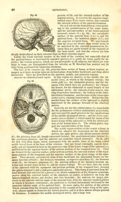
![the petrous portion exhibits a bony nucleus, stretching from its base towards its apex. The third point in order is that of the circle of the tympanum, a kind of ring channelled all round for the membrana tympani. This circle, at first almost horizontal, becomes gradually more and more oblique ; it is incomplete above, and the two extremities which are applied to the squamous portion cross each other instead of uniting. In many ani- mals the ring of the t}Tnpanum constitutes a distinct bone, named the tympanic hone. The fourth point of ossification appears in the mastoid portion during the fifth month. The last which becomes visible is that of the styloid process: it also remains distinct throughout life in the lower animals, and is called the styloid hone. It is not unconmion to find it in the same condition in the human subject. The development of these five pieces does not advance with equal rapidity. The petrous portion is most quickly completed. The mastoid, squamous, and petrous por- tions become united during the first year. The styloid process is attached to the rest of the bone at the age of two or three years ; at birth, the glenoid cavity is almost flat, on account of the absence of the auditory canal, and the shght development of the trans- verse root of the zygomatic process. The ulterior changes which take place in the temporal bone depend on the completion of the auditory canal and glenoid cavity, the increasing size of the mastoid process, and the obliteration of the projections and filling up of the hollows on the surface of the petrous portion. It is worthy of remark, that traces of the union of the base of the petrous portion with the squamous and mastoid portions, are visible in individuals of the most advan- ced age. The Cranium in general. The different bones which we have described unite in forming the cranium, an osse- ous cavity which encloses the brain, the cerebellum, and the annular protuberance. It is situated above the face, is the most elevated portion of the skeleton, and forms a con- tinuation of the vertebral column. The form of the cranium is that of an ovoid, flatten- ed below and at the sides, and with the large extremity turned backward. It is never perfectly symmetrical; but a very great deviation has always appeared to me coincident with disease of the brain. From attentive examination of a great number of sculls of idiots and maniacs, I have observed that in these subjects there is a remarkable differ- ence between the two sides. The dimensions of the cranium have been very accurately determined by Bichat. The antero-posterior diameter, measured from the foramen csecum to the occipital protuber- ance, is about five inches ;* the transverse diameter, measured between the base of the petrous portions of the temporal bones, is four inches and a half; the vertical diameter, extending from the anterior edge of the foramen magnum to the middle of the sagittal suture, is rather less than the transverse. In front, and behind the spot where the height and breadth of the cranium are measured, i. e., in front and behind the bases of the petrous bones, the diameters progressively diminish. Hence it follows, that the point where the cranium has the greatest capacity is the junction of the two anterior thirds with the posterior third ; that is to say, at the place of meeting, or, if I may use the expression, at the confluence of the brain, cerebellum, and spinal marrow. The cranium, however, presents many varieties, both in regard to its dimensions and shape. The varieties of form of the scull in different individuals appear generally to de- pend upon the preponderance of one diameter over another; and it may be remarked, that in these cases, where one diameter is much increased, the others are almost in- variably diminished in the same proportion, so that the absolute difference in size is by no means considerable. There are also variations in size and figure peculiar to the crania of different nations, as has been shown by the researches of Blumenbach and ScEnunerring. In the white, or Caucasian race, the cranium is decidedly much larger than in the others, more es- pecially than in the negro. Among certain tribes, the configuration of the cranium is determined by the permanent or frequently-repeated compressions to which the sculls of infants are subjected. It varies also according to age and sex, being proportionally larger in the fcetus than in the adult, and in the male than in the female. It should be remarked that all these varieties are exclusively confined to the vault of the cavity. Since the cranium is exactly moulded upon the brain, great interest has been attached to the exact appreciation of its dimensions, and hence the different measurements which have been adopted for this purpose. The oldest is the one proposed by Camper, under the name of the/ack/ angle. This angle is intended to measure the relative proportions of the cranium and face. It is taken by drawing one line from the middle incisors of the upper jaw along the front of the forehead, and another from the same point to the auditory meatus. The angle included between these lines is in the European from 80° to 85°, in the Mongolian race 75°, and in the negro, 70°. This anatomical fact had not escaped the attention of the ancients. We observe that in the statues of their heroes * [An old Paris inch is =1.065765 incli English.]](https://iiif.wellcomecollection.org/image/b21196801_0069.jp2/full/800%2C/0/default.jpg)