On the microscopic pathology of cancer, (with a woodcut) / by John Houston, M.D.
- John Houston
- Date:
- 1844
Licence: Public Domain Mark
Credit: On the microscopic pathology of cancer, (with a woodcut) / by John Houston, M.D. Source: Wellcome Collection.
Provider: This material has been provided by The University of Glasgow Library. The original may be consulted at The University of Glasgow Library.
10/20 (page 8)
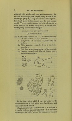
![sefies of cells are incased, one within the other, the smallest microscopic, the largest being visible to the nfakedeye. [Fig. 4.] They exist in extraordinary num- bers in all these instances, and are the real seminia morhi. The corpuscles seen through the microscope in their interior are either young cells, or nuclei from which young cellules are developed. EXPLANATION OF THE WOODCUT. (In part after Miiller.) iiG. 1. Plan of a primitive cell. a. The cell-membrane. 6. The nucleus, e. The nucleolus. 2. Cell-globules of carcinoma, magnified 500 dia- meters. 3. White granular corpuscles from a carcinoma mamillare. 4. Cells from a carcinoma areolare of the stomach. 5. Caudate corpuscles, of different shapes, from a carcinoma medullare. hi the observations which 1 have to make on the several varieties, I shall adopt the classification and nomenclature of Miiller. Carcinoma simplex The tumour of c. simplex is irregular in form, but not lobulated. It is hard and resisting to the knife (whence the term stone-cancer,)](https://iiif.wellcomecollection.org/image/b21476007_0010.jp2/full/800%2C/0/default.jpg)