Fistula in ano in phthisis, and actinomycosis bovis / by Heneage Gibbes.
- Gibbes, Heneage, -1912.
- Date:
- [1889]
Licence: Public Domain Mark
Credit: Fistula in ano in phthisis, and actinomycosis bovis / by Heneage Gibbes. Source: Wellcome Collection.
Provider: This material has been provided by The Royal College of Surgeons of England. The original may be consulted at The Royal College of Surgeons of England.
4/8 page 4
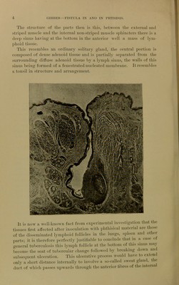
![The stnictnro of the piirts then is this, between the external jukI sti'i]ic(l muscle iuul the internal non-striped muscle sphincters there is a deep sinus having at the bottom in the anterior well a mass of lym- phoid tissue. This resembles an ordinary solitary gland, the central portion is composed of dense adenoid tissue and is partially separated from the suiTOunding dithise adenoid tissue by a lymph sinus, the walls of this sinus being formed of a fenestrated nucleated membrane. It resembles a tonsil in structure and arrangement. Piii It is now a well-known fact from experimental investigation that the tissues first affected after inoculation with phthisical material are those of the disseminated lymphoid follicles in the lungs, spleen and other parts; it is therefore perfectly justilial)le to conclude that in a case of general tuberculosis this lymph follicle at the bottom of this sinus may l,ecome the seat of tubercular change followed by breaking down and subsequent ulceration. This ulcerative process would have to extend only a short distance internally to involve a so-called sweat gland, tho duct of which passes upwards through the anterior fibres of the internal](https://iiif.wellcomecollection.org/image/b22301768_0006.jp2/full/800%2C/0/default.jpg)