Topographical anatomy of the dog / by O. Charnock Bradley.
- Orlando Charnock Bradley
- Date:
- 1927
Licence: In copyright
Credit: Topographical anatomy of the dog / by O. Charnock Bradley. Source: Wellcome Collection.
83/288 page 67
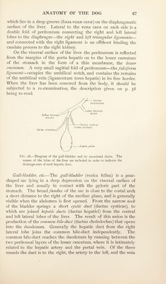
![which lies in a deep groove (fossa venae cavie) on the diaphragmatic surface of the liver. Lateral to the vena cava on each side is a double fold of peritoneum connecting the right and left lateral lobes to the diaphragm—the right and left triangular ligaments— and connected with the right ligament is an offshoot binding the caudate process to the right kidney. On the visceral surface of the liver the peritoneum is reflected from the margins of the porta hepatis on to the lesser curvature of the stomach in the form of a thin membrane, the lesser ornefitum. A very small sagittal fold of peritoneum—the falciform ligament—occupies the umbilical notch, and contains the remains of the umbilical vein (ligamentum teres hepatis) in its free border. When the liver has been removed from the body, it should be subjected to a re-examination, the description given on p. 38 being re-read. P’lG. 28.—Diagram of the gall-bladder and its associated ducts. The names of the lobes of the liver are included in order to indicate the drainage-area of each hepatic duct. Gall-hladder^ etc,—The gall-bladder (vesica fellea) is a pear- shaped sac lying in a deep depression on the visceral surface of the liver and usually in contact with the pyloric part of the stomach. The broad fundus of the sac is close to the costal arch a short distance to the right of the median plane, and is generally visible when the abdomen is first opened. From the narrow neck of the bladder springs a short cystic duct (ductus cysticus), to which are joined hepatic ducts (ductus hepatici) from the central and left lateral lobes of the liver. The result of this union is the production of the common bile-duct (ductus choledochus) that opens into the duodenum. Generally the hepatic duct from the right lateral lobe joins the common bile-duct independently. The common bile-duct reaches the duodenum by running between the two peritoneal layers of the lesser omentum, where it is intimate])^ related to the hepatic artery and the portal vein. Of the three vessels the duct is to the right, the artery to the left, and the vein](https://iiif.wellcomecollection.org/image/b29820108_0083.jp2/full/800%2C/0/default.jpg)
 No text description is available for this image
 No text description is available for this image
 No text description is available for this image