The immune system: genes, receptors, signals : [proceedings] / edited by Eli E. Sercarz, Alan R. Williamson [and] C. Fred Fox.
- ICN-UCLA Symposium on Molecular Biology
- Date:
- 1974
Licence: Attribution-NonCommercial 4.0 International (CC BY-NC 4.0)
Credit: The immune system: genes, receptors, signals : [proceedings] / edited by Eli E. Sercarz, Alan R. Williamson [and] C. Fred Fox. Source: Wellcome Collection.
644/664 page 616
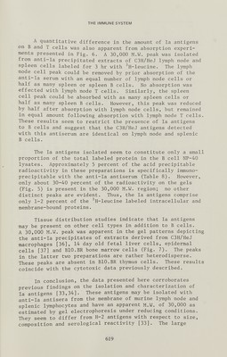
![H. о. McDEVITTefa/. terize the related functional membrane components [3,33,34]. Several attempts to identify the presently known la alloantigenic specificities on different lymphoid cells, using cytotoxic and adsorption tests performed with anti-la antisera raised in ^ congenie recombinant strains, have been reported [5,23,24,27,28]. These assays unexpectedly revealed that la antigens can be identified primarily on lymph node and splenic В cells. However, they may be pres¬ ent, but in much lower amount on peripheral T cells. An initial biochemical characterization of these cellular anti¬ gens examined by SDS-polyacrylamide gel electrophoresis of radiolabeled, NP-40 solubilized and anti-la precipitated membrane-associated proteins demonstrated them to have a M.W. of approximately 30,000 daltons, under reducing condi¬ tions, thus differing in size from H-2 antigens [33,34]. This type of analysis has been extended by determining whether these proteins could be isolated from membranes of T and В cells, as well as other cell types. Polyacrylamide gel electrophoretic patterns of reduced immune precipitates formed between an (A.TH x BlO.S) anti- (A.TL x BlO.S) antiserum and NP-40 extracts [33,34] of ^H- leucine labeled splenic Б cells and lymph node T cells pre¬ pared [5] from mice of several H-2 haplotypes are shown in Fig. 5. These profiles indicate that membrane proteins of approximately 30,000 M.W. are isolatable by this method from В cells and not from T cells. No such peaks of radio¬ activity were obtained in the control precipitates of either cell type using normal mouse serum. Thus, the cellu¬ lar and strain distributions of these la antigens as well as their size heterogeneity correlate closely with the pattern of reactivity reflected by the cytotoxic assays previously described (see Table 7). This antiserum reacts predominant¬ ly with В cells of Ir^, cross-reacts with В cells which are Ir*^ and Ir^, but shows no reactivity with В cells of Ir s (Fig. 5 and Table 8). It should be noted that while the anti-la serum does not react with purified T cells, anti-K^ and anti-K® antisera do react with the appropriate T cells (Table 8). In other experiments (Delovitch, unpublished data), a congenie anti-Thy 1.2 antiserum precipitates about 1-2 percent of the label in the T cell lysates. These results suggest that В cells definitely possess la antigens. While these antigens are not detectable on T cells under the conditions employed, it is possible that they are present in reduced quantities on T cells. 616](https://iiif.wellcomecollection.org/image/b18036387_0645.JP2/full/800%2C/0/default.jpg)


 No text description is available for this image
 No text description is available for this image
 No text description is available for this image