The immune system: genes, receptors, signals : [proceedings] / edited by Eli E. Sercarz, Alan R. Williamson [and] C. Fred Fox.
- ICN-UCLA Symposium on Molecular Biology
- Date:
- 1974
Licence: Attribution-NonCommercial 4.0 International (CC BY-NC 4.0)
Credit: The immune system: genes, receptors, signals : [proceedings] / edited by Eli E. Sercarz, Alan R. Williamson [and] C. Fred Fox. Source: Wellcome Collection.
647/664 page 619
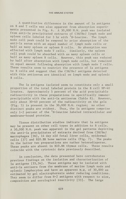
![THE IMMUNE SYSTEM A quantitative difference in the amount of la antigens on В and T cells was also apparent from absorption experi¬ ments presented in Fig. 6. A 30,000 M.W. peak was isolated from anti-la precipitated extracts of C3H/HeJ lymph node and spleen cells labeled for 3 hr with ^H-leucine. The 1зтпрЬ node cell peak could be removed by prior absorption of the ^^ti-Ia serum with an equal number of lymph node cells or half as many spleen or spleen В cells. No absorption was effected with Ijmiph node T cells. Similarly, the spleen cell peak could be absorbed with as many spleen cells or half as many spleen В cells. However, this peak was reduced by half after absorption with lymph node cells, but remained in equal amount following absorption with lymph node T cells. These results seem to restrict the presence of la antigens to В cells and suggest that the C3H/HeJ antigens detected with this antiserum are identical on lymph node and splenic В cells. The la antigens isolated seem to constitute only a small proportion of the total labeled protein in the В cell NP-40 lysates. Approximately 5 percent of the acid precipitable radioactivity in these preparations is specifically immuno- precipitable with the anti-la antiserum (Table 8). However, only about 30-40 percent of the radioactivity on the gels (Fig. 5) is present in the 30,000 M.W. region; no other distinct peaks are evident. Thus, the la antigens comprise only 1-2 percent of the ^H-leucine labeled intracellular and membrane-bound proteins. Tissue distribution studies indicate that la antigens may be present on other cell types in addition to В cells. A 30,000 M.W. peak was apparent in the gel patterns depicting the anti-la precipitates of extracts derived from СЗН/HeJ macrophages [36], 14 day old fetal liver cells, epidermal cells [37] and BIO.BR bone marrow cells (Fig. 7). The peaks in the latter two preparations are rather heterodisperse. These peaks are absent in BIO.BR thymus cells. These results coincide with the cytotoxic data previously described. In conclusion, the data presented here corroborates previous findings on the isolation and characterization of la antigens [33,34]. These antigens may be isolated with anti-la antisera from the membrane of murine lymph node and splenic lymphocytes and have an apparent M.W. of 30,000 as estimated by gel electrophoresis under reducing conditions. They seem to differ from H-2 antigens with respect to size, composition and serological reactivity [33]. The large 619](https://iiif.wellcomecollection.org/image/b18036387_0648.JP2/full/800%2C/0/default.jpg)


 No text description is available for this image
 No text description is available for this image
 No text description is available for this image