The brain of the cat (Felis domestica). 1. Preliminary account of the gross anatomy / by Burt G. Wilder.
- Burt Green Wilder
- Date:
- [1881]
Licence: Public Domain Mark
Credit: The brain of the cat (Felis domestica). 1. Preliminary account of the gross anatomy / by Burt G. Wilder. Source: Wellcome Collection.
Provider: This material has been provided by The Royal College of Surgeons of England. The original may be consulted at The Royal College of Surgeons of England.
27/48 page 549
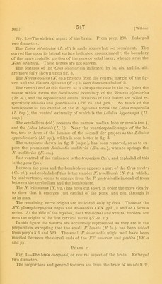
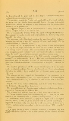
![1881.] the less extent of the pons, and the less degree of lle.xurc of the whole brain at the mesenceiihalic region. The greater width of the Tractus postrliinalis (Tr. prh.), which includes the surface of the Lobulus hypocnmpos {LI, In man, indeed, this part is hardly visible on account of the prominence of the convolutions laterad of the F. postrhmalis. The apparent origin of the F. oculomotorius {N. ocm.) laterad of the meson, and just caudad of the cimbia {c7nb.). The appearance of a division of the ectal layers of the pontile fibres into three groups, cephalic, caudal and intermediate, the latter partly over- lapped by the other two. The appearance of a faint band crossing the trapezium a little obliquely between the origins of the NF. abdmens {N. abd.) and facialis {N. /.). The distinctness of this band varies. The origin of the N. hypoglossus {N. hg.) laterad of the Area elUptka {Ar. el.), which might otherwise be taken as the surfiice of the olimi or “ olivary body ” of man. The determination of this point involves some comparisons and sections which I have not yet made, so I merely in- dicate the part by a descriptive name and leave the question open. The close association of the roots of the AfAf. glossopharyiigeus (iV. gpli.), 'oagus {N. «.), and accessorius {N. ac.). The long caudal nerve is of course accessorial, and the cephalic funiculi are unquestionably glossopharyn- geal ; but how the intermediate funiculi should be assigned, I am not yet sure.* The marked prominence of the ventro-lateral region of the metence- phalic continuation of the Golumna lateralis myelonis {Glin. 1.), forming an elevation to which I have applied the provisional name Ai'ea ooalis {Ar. ov.). The absence of any superficial decussation of the pyramids (;»/.). Hence, the F. ventrimesalis {F. vms.) or “anterior fissure,’’ is uninterrupted. The F. ventrUateralis {F. vl.) is deflected at the caudal end of the Area elliptica. Fig. 4.—The raesal surface of the right half of the brain (hemiencepha- lon dextruui). Enlarged two diameters. The general features are from the same brain as fig. 3, but some features are derived from prep’s 290, 304 and 454. The surfaces shown in tliis figure are of four kinds, as follows ;— 1. The natural surfaces wliich are covered b}”^ pia. Tliese are the mesal aspects of the hemisphere {hem.), and the Lobus olfaciorius {L. ol.). 2. The natural mesal surface {Ar. spt.) of the right half of the septum lucidum, which, in the cat, is in contact with its lateral homologue, or separated tlierefrom only by a thin layer of connective. I have never observed an interval corresponding to the pseudocidia or “liflli ventricle’’ of man. •In a paper on the N. vagus In tlie cal, proscnlcil at tl\c same time with this, Prof. T. B. Stowell has given a fuller aecouut of the relations of these nerves.](https://iiif.wellcomecollection.org/image/b22381983_0029.jp2/full/800%2C/0/default.jpg)


 No text description is available for this image
 No text description is available for this image
 No text description is available for this image