A manual of diseases of the throat and nose / by Francke Huntington Bosworth.
- Date:
- 1881
Licence: Public Domain Mark
Credit: A manual of diseases of the throat and nose / by Francke Huntington Bosworth. Source: Wellcome Collection.
Provider: This material has been provided by the Royal College of Physicians of Edinburgh. The original may be consulted at the Royal College of Physicians of Edinburgh.
102/460 (page 76)
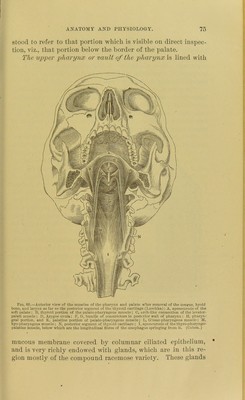
![7G aie aggregated to such an extent as to give it the name of the pharyngeal tonsil. They secrete an abundant mucus, which is poured out upon the membrane and serves to keep it in a soft, moist, and pliable condition. It is also squeezed out as it were in the act of deglutition, and serves to coat the bolus of food, thus facilitating its passage down the oesophagus. The pharynx proper, or lower pharynx, viz., that 'portion below the border of the palate, is lined with mucous membrane more closely adherent to the parts than that of the upper pharynx. It is covered with squamous epithelium and is en- dowed with simple and compound follicular glands, somewhat sparsely scattered through the membrane. The soft palate is a movable fold of membrane which is sus- pended from the posterior border of the hard palate. It contains a number of muscles by. which it is acted upon, and is endow- ed with certain functions in con- nection with articulation and phonation, and also in the act of deglutition. With its ac- tion in the formation of the voice we need not concern our- selves further than to say that by its position, in partially or completely closing the nasal cavity posteriorly, it modifies the character of the voice, giv- ing it or depriving it of its na- sal tone. In deglutition the palate is drawn firmly backward and upward against the posterior wall of the pharynx, complete- ]y closing the opening between the mouth and posterior nares, thus preventing the entrance of the bolus into the nasal cavity. This is mainly accomplished by the contraction of the palato- pliaryngeus muscle, which forms almost a complete circular muscle resembling the orbicularis. This is shown in Fig. 68, which illustrates the distribution of muscles on the under sur- Pig. 69.—Anterior view of the naso-pharyngeal space; on one side the mncons membrane has bi en dissected away (after Lnschka). 3, septum; 2, middle; 3, lower turbinated bone; 4. tuberosity of the pharyngeal orifice of the Eustachian tube; 5. soft palate ; 6, uvula; 7, stylo-pharyngeus mns- cie; 8, levator-palati; !), palato-pharyngeus mus- cle. (Ziemssen.)](https://iiif.wellcomecollection.org/image/b21932165_0102.jp2/full/800%2C/0/default.jpg)