On abnormal nutrition in articular cartilages / by P. Redfern, M.D.
- Redfern, Peter, 1821-1912
- Date:
- 1849
Licence: Public Domain Mark
Credit: On abnormal nutrition in articular cartilages / by P. Redfern, M.D. Source: Wellcome Collection.
Provider: This material has been provided by The University of Glasgow Library. The original may be consulted at The University of Glasgow Library.
42/90 page 40
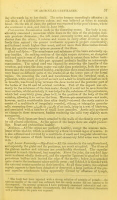
![deeper parts, it is dark and molecular, or indistinctly fibrous, the fibres running perpendicularly to the surface. The yellow spot on the cartilage of the head of the femur is found to be owing to the existence of a quantity of oil in the substance of the tissue ; but no fatty matter exists within the cells, though they are much enlarged. In some places, where the cartilage is not more than :^th of an inch thick, the supei-ficial cells are not all removed, and yet no progressive enlargement of the deep cells is ob- served on passing down to the bone, as it is on passing towards tlie free surface at jmrts where that constitutes the principal or sole point of absorption. On examining the edge of the cartilage at the margin of the surfaces, and near to the attachment of the round ligament, after stripping off the fibrous membrane from it, the hyaline substance is found to be converted into distinct interlacing fibres, amongst which are numbers of cells or corpuscles, the whole tissue at these parts being almost precisely similar to fibro-cartilage. The microscopic appearances in the diseased parts of the other cartilages of these extremities are similar to those above-named. Where there is a fibrous membrane on the surface, or where fibrous processes project from it, these con- sist of bands or fibres running in different directions according to the position of the cells where the fibres are formed. Amongst the fibres are a few cartil- age cells, or granular patches indicating their former existence, and always seen after the action of acetic acid. The mode of junction of the fibrous layer, found on the surface, with the cartilage, and the changes which are seen to have taken place in the superficial and deep cells are precisely similar to those which have been already described in the cartilages of the left femur and acetabulum. Upper Extremities.—The cartilage covering the heads of both humeri pre- sents whitish patches, as if there were a deposit of dense matter immediately below the surface. The cartilage of the glenoid cavities is healthy. Elbow-Joint.—The cartilage on the lower articular surfaces of the humeri is reddened and velvetty on the small head and the adjacent ridge,—that on the right humerus presents an abraded spot on the trochlear surface, and an elongated but deep notch, ^th of an inch long, and ^Vth broad, at the anterior part of the ridge, separating the trochlear surface from the small head. The cartilage on the heads of the radii is reddened and velvetty or fib- rous, especially at the edge of the humeral surface, and at the part near to the ulna, where nearly the whole thickness of the layer is diseased. The cartilage on the ulna is slightly reddened and velvetty. On each side, it is divided into two parts by an irregular membranous band passing across at the junction of the olecranon with the shaft. At this point, the bone ap- pears almost bare, except at the radial end of the band on the left ulna, where there is still a layer of cartilage. Wrist-Joints.left wrist-joint is healthy. In the right one the cartil- ages covering the lower surfaces of the radius and ulna, are converted into fibres at their margins, whilst their central parts are covered by a thin membrane. Right-Hand.—Many parts of the cartilages of the bones in the lower carpal row are reddened, velvetty, and abraded. The semilunar surface of the unci- form, and the unciform surface of the fourth metacarpal bone, present irre- gular spots, where the bone is completely bare, smooth, and covered with por- cellanous deposit. Left-Hand.—The cartilage on the lower surface of the sca])hoid, and on the semilunar surface of the unciform bone, is removed on each for an extent of 1-Gth of an inch. The exposed bone is perfectly smooth and porcellan- ous, and the edge of tiie surrounding cartilage is loose and velvetty. The surface of the trapezoid for the trapezium is in the same state. Several other cartilages of the carpal bones are reddened and velvetty, and especially the cartilages in the carpo-mctacarpal articulation of the thumb. Microscopic Examination.—On examining vertical sections of the cartilage of tlie heads of the humeri, passing through the wliite spots, these are found to be](https://iiif.wellcomecollection.org/image/b21470704_0042.jp2/full/800%2C/0/default.jpg)


 No text description is available for this image
 No text description is available for this image
 No text description is available for this image