The microscope in its application to practical medicine / by Lionel S. Beale.
- Lionel Smith Beale
- Date:
- MDCCCLXVII [1867]
Licence: Public Domain Mark
Credit: The microscope in its application to practical medicine / by Lionel S. Beale. Source: Wellcome Collection.
Provider: This material has been provided by King’s College London. The original may be consulted at King’s College London.
55/510
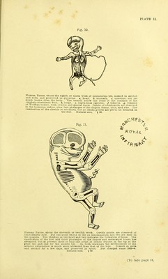
![PLATE II. Fig. 10. Human Fcetus, about the eighth or ninth week of intrauterine life, soaked in alcohol and soda, and preserved in glycerine, a. heart, b, stomach, c. intestine, not yet much longer than the body. The branch below the letter is the remains of the °?1Ptia]°'mesenteric duct, d, lungs, e. supra-renal capsules. /, kidneys, g. remains oi Wolffian bodies, with ovaries and genital ducts. Points of ossification are observed in the humerus, radius, ulna, last phalanges of the fingers, femur, tibia, and ribs. The ossification of the clavicle is advanced, but no ossific points are yet to be detected in the feet. Natural size. $ 90. Human Foetus, about the eleventh or twelfth week. Ossific points are observed of considerable size. But one point exists in the os innominatum and two are seen in the scapula. The shading in the head and face indicates the formation of bone. The ossification of the first and third phalanges of the fingers and metacarpal bones has advanced, but at present there is only one point of ossific deposit in the tip of the great toe and one for the middle toe. In both drawings the development of the anterior extremities is much more advanced than that of the legs. Soaked in soda and alcohol for a few days, and preserved in spirit. Not changed since 1853—4. Natural size. § 90. [To face page 24.](https://iiif.wellcomecollection.org/image/b21302492_0055.jp2/full/800%2C/0/default.jpg)