Note on the bodies recently described by Leishman and Donovan / by R. Ross.
- Ronald Ross
- Date:
- 1903
Licence: Public Domain Mark
Credit: Note on the bodies recently described by Leishman and Donovan / by R. Ross. Source: Wellcome Collection.
4/6 page 4
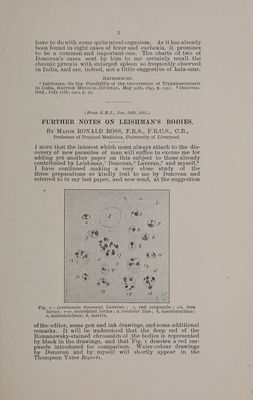
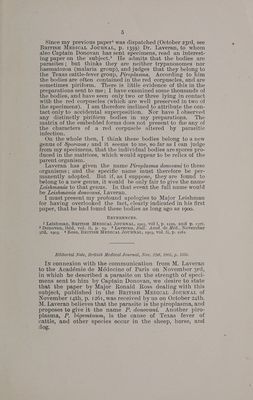
![It seems to me very important to note that in the prepara- tions made intra wtam Leishman’s bodies appear under two distinct conditions, namely, (a) free, and (6) embedded, to the number of one up to twelve, in a matrix. Figs. 2 to 6 illus- trate the free forms.. In the majority the contour is elliptic, but it is occasionally nearly circular. It is necessary to re- cognize that the contour line is a strong one, suggestive of a well-marked cell wall (that is, in the etra-vitam specimens, which are, of course, the most suitable for study). The two chromatin masses are generally situated at the extremities of the minor diameter of the elliptic cell (Figs. 2 to5), The larger mass often seems to bulge beyond the cell wall (Figs. 2to 4). I think that both masses are always present, but that the smaller one may be occasionally hidden by the larger. In my specimens the protoplasm of the cell is a faint red (with- out tinge of blue), and becomes fainter from the contour in- wards, so that there is the characteristic clear area round the macronucleus. In short, all the free forms have a most definite size and structure. Much interest, however, attaches to those bodies which are embedded in a matrix (Figs. 7 to 17). These are found only in the preparations made intra wtam, and are much less numerous than the free forms. Moreover the contour of the little cell is generally much less distinct in the embedded organisms than in the free ones; although the chromatin is stained just as deeply and the two chromatin masses bear just the same relations to each other as regards position and distance. What I eall (until a more definite term can be employed) the matrix is always stained very faintly in these specimens—much more faintly than the red or white cor- puscles. The tint is violet, or more rarely mauve. The structure appears cloudy, or perhaps granular, or even stromatic; but is too delicate for expression in the drawings. The form is generally a more or less regular oval (Figs. 8 to 16); but sometimes the mass appears to be shapeless or torn (Figs. 7 to 17). The outline is sharp; but it is important to note that there is never any contour line suggestive of a cell- wall, as with the Leishman bodies themselves. There is never, also, any suggestion of the haemoglobin of the red corpuscles, or the nucleus of the white corpuscles, to be seen in this matrix. Its size varies in my specimens from about 3 to 8u in the long diameter; and there is a rough, but only a rough, correspondence between the size of the matrix and the number of Leishman bodies it contains. Sometimes, as in Fig. 13, a considerable part of the matrix is empty; and sometimes, a rather large matrix holds only one of the bodies. Fig. 17 shows eight bodies in one matrix: but I have seen as many as twelve. Occasionally we find clusters of the bodies touching each other, without lying in any visible matrix ; but in such cases their oval contours are well marked. One or more of the micronuclei are sometimes not to be seen. I should add that Donovan clearly shows, in his letter and drawings sent to me, that he has well observed all these forms, In his recent paper’ Leishmann stil] inclines to the view that these bodies are “‘altered trypanosomes.” In that case it is singular that in two whole specimens made during life and containing large numbers of them, we should not find a single unaltered trypanosome, or even the flagellum of one. I may be wrong, but I can see little in these objeets to reeall the involution forms of trypanosomes.](https://iiif.wellcomecollection.org/image/b33627757_0004.jp2/full/800%2C/0/default.jpg)