Text-book of normal histology : including an account of the development of the tissues and of the organs / By George A. Piersol ... With four hundred and nine illustrations, of which three hundred and fifty-eight are from original drawings by the author.
- George A. Piersol
- Date:
- 1899
Licence: Public Domain Mark
Credit: Text-book of normal histology : including an account of the development of the tissues and of the organs / By George A. Piersol ... With four hundred and nine illustrations, of which three hundred and fifty-eight are from original drawings by the author. Source: Wellcome Collection.
Provider: This material has been provided by the Augustus C. Long Health Sciences Library at Columbia University and Columbia University Libraries/Information Services, through the Medical Heritage Library. The original may be consulted at the the Augustus C. Long Health Sciences Library at Columbia University and Columbia University.
15/456 page 11
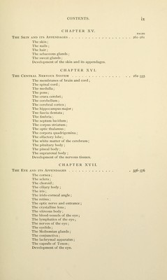
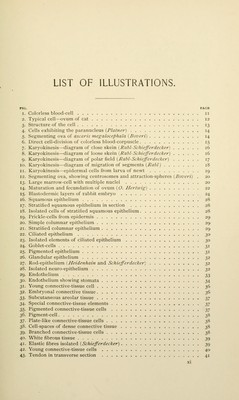
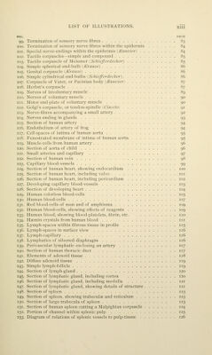
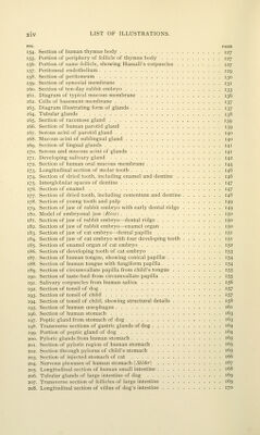
![LIST OF ILLUSTRATIONS. FIG. PAGB 1. Colorless blood-cell ii 2. Typical cell—ovum of cat 12 3. Structure of the cell 13 4. Cells exhibiting the paranucleus {Plainer) 14 5. Segmenting ova of ascaris niegalocephala {Boveri) 14 6. Direct cell-division of colorless blood-corpuscle 15 7. Karyokinesis—diagram of close skein (Rabl-Schiefferdecker) 16 8. Karyokinesis—diagram of loose skein \ Rabl-Schiefferdecker) 16 9. Karyokinesis—diagram of polar field {Rabl-Schiefferdecker) 17 10. Karyokinesis—diagram of migration of segments {Rabl) 18 11. Karyokinesis—epidermal cells from larva of newt 19 12. Segmenting ova, showing centrosomes and attraction-spheres {Boveri) . 20 13. Large marrow-cell with multiple nuclei 20 14. Maturation and fecundation of ovum (O. Hertwig] 22 15. Blastodermic layers of rabbit embryo 24 16. Squamous epithelium 28 17. Stratified squamous epithelium in section 28 18. Isolated cells of stratified squamous epithelium 28 19. Prickle-cells from epidermis 29 20. Simple columnar epithelium 29 21. Stratified columnar epithelium 29 22. Ciliated epithelium Tp 23. Isolated elements of ciliated epithelium 30 24. Goblet-cells 31 25. Pigmented epithelium 31 26. Glandular epithelium 32 27. Rod-epithelium (Heidenhain and Schiefferdecker) 32 28. Isolated neuro-epithelium 32 29. Endothelium t^t, 30. Endothelium showing stomata 34 31. Young connective-tissue cell 36 32. Embryonal connective tissue 36 2,2). Subcutaneous areolar tissue t,-j 34. Special connective-tissue elements 2>7 35. Pigmented connective-tissue cells yj 36. Pigment-cell 38 37. Plate-like connective-tissue cells 38 38. Cell-spaces of dense connective tissue 38 39. Branched connective-tissue cells t^^ 40. White fibrous tissue 39 41. Elastic fibres isolated {Schiefferdecker) 39 42. Young connective-tissue cells 40 43. Tendon in transverse section 41](https://iiif.wellcomecollection.org/image/b21210809_0015.jp2/full/800%2C/0/default.jpg)
 No text description is available for this image
 No text description is available for this image
 No text description is available for this image