Text-book of normal histology : including an account of the development of the tissues and of the organs / By George A. Piersol ... With four hundred and nine illustrations, of which three hundred and fifty-eight are from original drawings by the author.
- George A. Piersol
- Date:
- 1899
Licence: Public Domain Mark
Credit: Text-book of normal histology : including an account of the development of the tissues and of the organs / By George A. Piersol ... With four hundred and nine illustrations, of which three hundred and fifty-eight are from original drawings by the author. Source: Wellcome Collection.
Provider: This material has been provided by the Augustus C. Long Health Sciences Library at Columbia University and Columbia University Libraries/Information Services, through the Medical Heritage Library. The original may be consulted at the the Augustus C. Long Health Sciences Library at Columbia University and Columbia University.
17/456 page 13
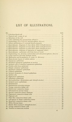
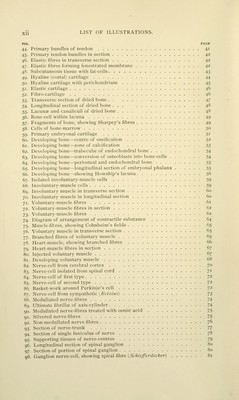
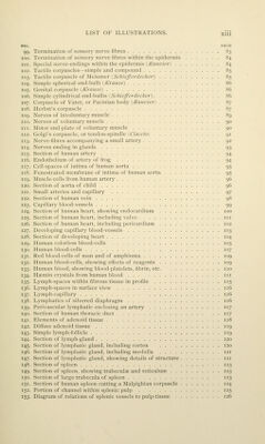
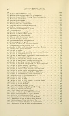
![PIG. PAGE 99. Termination uf sensory nerve fibres 83 100. Termination of sensory nerve fibres within the ejiidermis 84 loi. Special nerve-endings within the epidermis I/v(Z«c'/Vrj 84 102. Tactile corpuscles—simple and com]K)nnd 85 103. Tactile corpuscle of .Mcissner I .SW//V^V;7/<'f^<';-) 85 104. Simple spherical end-bulb (Krause) 86 105. Genital corpuscle ^A'rause) 86 106. Simple cylindrical end-bulbs 15c/^iV:^<?;'rffrA^r) ... 86 107. Corpuscle of Vater, or Pacinian body (Ranvier) 87 108. Herbst's corpuscle 87 109. Nerves of involuntary muscle 89 110. Nerves of voluntary muscle 90 111. Motor end-plate of voluntary muscle 90 112. Golgi's corpuscle, or tendon-spindle (Ci'atczo) 91 113. Nerve-fibres accompanying a small artery 92 114. Nerves ending in glands 93 115. Section of human artery 94 116. Endothelium of artery of frog 94 117. Cell-spaces of intima of human aorta 95 118. Fenestrated membrane of intima of human aorta 95 119. Muscle-cells from human artery 96 120. Section of aorta of child 96 121. Small arteries and capillary 97 122. Section of human vein 98 123. Capillar)' blood-vessels 99 124. Section of human heart, showing endocardium 100 125. Section of human heart, including valve 101 126. Section of human heart, including pericardium 102 127. Developing capillary blood-vessels 103 128. Section of developing heart 104 129. Human colorless blood-cells 105 130. Human blood-cells 107 131. Red blood-cells of man and of amphiuma 109 132. Human blood-cells, showing effects of reagents 109 133. Human blood, showing blood-platelets, fibrin, etc no 134. Haemin crystals from human blood in 135. Lymph-spaces within fibrous tissue in profile 115 136. Lj'mph-spaces in surface view 116 137. Lymph-capillary 116 138. Lymphatics of silvered diaphragm ii5 139. Perivascular l\-mphatic enclosing an artery 117 140. Section of human thoracic duct 117 141. Elements of adenoid tissue 118 142. Diffuse adenoid tissue 119 143. Simple lymph-follicle 119 144. Section of lymph-gland 120 145. Section of lymphatic gland, including cortex 120 146. Section of h^mphatic gland, including medulla 121 147. Section of lymphatic gland, showing details of structure 121 148. Section of spleen 123 149. Section of spleen, showing trabeculae and reticulum 123 150. Section of large trabecula of spleen 123 151. Section of human spleen cutting a Malpighian corpuscle 124 152. Portion of channel within splenic pulp 125 153. Diagram of relations of splenic vessels to pulp-tissue 126](https://iiif.wellcomecollection.org/image/b21210809_0017.jp2/full/800%2C/0/default.jpg)
 No text description is available for this image
 No text description is available for this image
 No text description is available for this image