Text-book of normal histology : including an account of the development of the tissues and of the organs / By George A. Piersol ... With four hundred and nine illustrations, of which three hundred and fifty-eight are from original drawings by the author.
- George A. Piersol
- Date:
- 1899
Licence: Public Domain Mark
Credit: Text-book of normal histology : including an account of the development of the tissues and of the organs / By George A. Piersol ... With four hundred and nine illustrations, of which three hundred and fifty-eight are from original drawings by the author. Source: Wellcome Collection.
Provider: This material has been provided by the Augustus C. Long Health Sciences Library at Columbia University and Columbia University Libraries/Information Services, through the Medical Heritage Library. The original may be consulted at the the Augustus C. Long Health Sciences Library at Columbia University and Columbia University.
22/456 page 18
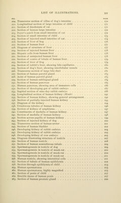
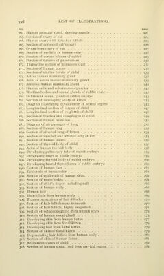
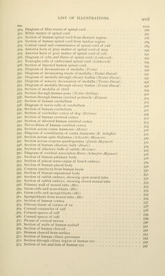
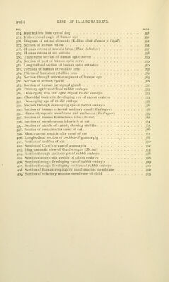
![PIG. PAGE 374. Injected iris from eye of dog 348 375. Irido-corneal angle of human eye 350 376. Diagram of retinal elements (Kallius after Ramony Cajal) 352 377. Section of human retina 353 378. Human retina at macula lutea (Max Schullze) 357 379. Human retina at ora serrata 358 380. Transverse section of human optic nerve 359 381. Section of part of human optic nerve 359 382. Longitudinal section of human optic entrance 360 383. Portions of human crystalline lens 362 384. Fibres of human crystalline lens 362 385. Section through anterior segment of human eye 363 386. Section of human eyelid 368 387. Section of human lachrymal gland 371 388. Primary optic vesicle of rabbit embryo t;jt, 389. Developing lens and optic cup of rabbit embryo 373 390. Choroidal fissure in developing eye of rabbit embryo T)-]-^ 391. Developing eye of rabbit embryo 375 392. Section through developing eye of rabbit embryo 376 393. Section of human external auditory canal ^Riidinger) 378 394. Human tympanic membrane and malleolus [Riidinger) 379 395. Section of human Eustachian tube ( Tesiut) 382 396. Section of membranous labyrinth of cat 384 397. Section of utricle of rabbit, showing otoliths 385 398. Section of semicircular canal of cat 386 399. Membranous semicircular canal of cat 387 400. Longitudinal section of cochlea of guinea-pig , 388 401. Section of cochlea of cat 390 402. Section of Corti's organ of guinea-pig 392 403. Diagrammatic view of Corti's organ ( Testul) 393 404. Section through auditory pit of rabbit embryo 398 405. Section through otic vesicle of rabbit embryo 398 406. Section through developing ear of rabbit embrj-o 399 407. Section through developing cochlea of rabbit embryo 400 408. Section of human respiratory nasal mucous membrane 402 40^ Section of olfactory mucous membrane of child 403](https://iiif.wellcomecollection.org/image/b21210809_0022.jp2/full/800%2C/0/default.jpg)
 No text description is available for this image
 No text description is available for this image
 No text description is available for this image