Atlas of the descriptive anatomy of the human body / by J.C. Cruveilhier ; with explanations by C. Bonamy; the figures drawn from nature by Émile Beau.
- Bonamy, Constantin Louis, 1812-
- Date:
- 1844
Licence: Public Domain Mark
Credit: Atlas of the descriptive anatomy of the human body / by J.C. Cruveilhier ; with explanations by C. Bonamy; the figures drawn from nature by Émile Beau. Source: Wellcome Collection.
Provider: This material has been provided by The University of Glasgow Library. The original may be consulted at The University of Glasgow Library.
76/284
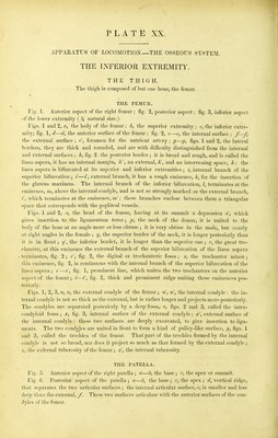
![APPARATUS OF LOCOMOTION—THE OSSEOUS SYSTEM. THE INFERIOR EXTREMITY. THE THIGH. The thigh is composed of but one bone, the femur. THE FEMUR. Fig. 1. Anterior aspect of the right femur ; fig. 2, posterior aspect; fig. 3, inferior aspect of the lower extremity ( % natural size.) Figs. 1 and 2, a, the body of the femur ; b, the superior extremity ; c, the inferior extre- mity; fig. 1, d—d, the anterior surface of the femur ; fig. 2, e—e, the internal suiface ; /—/, the external surface ; e', foramen for the nutrient artery ; ^—ff, figs. 1 and % the lateral borders, they are thick and rounded, and are with difficulty distinguished from the internal and external surfaces ; k, fig. 2. the posterior border ; it is broad and rough, and is called the linea aspera, it has an internal margin. A, an external, k', and an intervening space, k: the linea aspera is bifurcated at its superior and inferior extremities ; t, internal branch of the superior bifurcation ; i'—i', external branch, it has a rough eminence, k, for the insertion of the gluteus maximus. The internal branch of the inferior bifurcation, /, terminates at the eminence, m, above the internal condyle, and is not so strongly marked as the external branch, /', which terminates at the eminence, m'; these branches enclose between them a triangular space that corresponds with the popliteal vessels. Figs. 1 and 2, o, the head of the femur, having at its summit a depression o', which gives insertion to the ligamentum teres ; j?, the neck of the femur, it is united to the body of the bone at an angle more or less obtuse ; it is very obtuse in the male, but nearly at right angles in the female ; q, the superior border of the neck, it is longer posteriorly than it is in front; q', the inferior border, it is longer than the superior one ; ;•, the great tro- chanter, at this eminence the external branch of the superior bifurcation of the linea aspera terminates, fig. 2 ; fig. 2, the digital or trochanteric fossa ; s, the trochanter minor ; this eminence, fig. 2, is continuous with the internal branch of the superior bifurcation of the linea aspera ; s'—s', fig. 1, prominent line, which unites the two trochanters on the anterior aspect of the femur; t—I', fig. 2, thick and prominent ridge uniting these eminences pos- teriorly. Figs. 1, 2, 3, u, u, the external condyle of the femur ; u', ti', the internal condyle : the in- ternal condyle is not so thick as the external, but is rather longer and projects more posteriorly. The condyles are separated posteriorly by a deep fossa, v, figs. 2 and 3, called the inter- condyloid fossa ; x, fig. 3, internal surface of the external condyle ; x', external surface of tlie internal condyle : these two surfaces are deeply excavated, to give insertion to liga- ments. The two condyles are united in front to form a kind of pulley-like surfece, figs. 1 and 3, called the trochlea of the femur. That part of the trochlea formed by the internal condyle is not so broad, nor does it project so much as that formed by the external condyle ; z, the exfernal tuberosity of the femur ; z', the internal tuberosity. THE PATELLA. Fig. 5. Anterior aspect of the right patella ; a—b, the base ; c, the apex or summit. Fig. 6. Posterior aspect of the patella; a—b, the base ; c, the apex ; d, vertical ridge, that separates the two articular surfaces ; the internal articular surface, e, is smaller and less deep than the external, J] These two surfaces articulate with the anterior surfaces of the con- dyles of the femur.](https://iiif.wellcomecollection.org/image/b21458911_0076.jp2/full/800%2C/0/default.jpg)


 No text description is available for this image
 No text description is available for this image
 No text description is available for this image